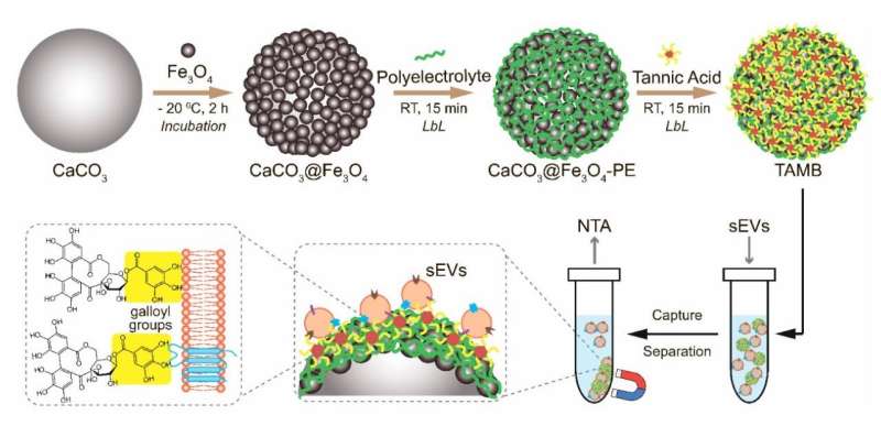
Formation of the shell of magnetic microparticles and the principle of their interaction with exosomes. Credit: Grishaev et al.
Researchers have synthesized tannic acid-coated magnetic beads capable of extracting membrane vesicles called "exosomes" from biological fluids with 60% efficiency. This novel method will make it easier and faster to isolate exosomes from laboratory samples, while the chemical composition of the exosomes will help detect cancer in its early stages. The study was published in the Journal of Materials Chemistry B.
Cells in the human body "communicate" through exosomes—tiny membrane vesicles that are released into the intercellular space and carry proteins, fats, nucleic acids, and other molecules. Since the exosomes' contents replicate the chemical composition of the cell they split off from, they can be used as molecular markers to identify various diseases. For example, the presence in exosomes of proteins and RNA typical of cancer cells is compelling evidence of incipient cancer.
However, exosome-based diagnostics is not yet widely used in clinical practice because these vesicles are as small as viruses and therefore difficult to isolate. A recently proposed approach uses magnetic beads to capture exosomes from biological samples, such as blood.
The surface of the beads is coated with molecules, such as antibodies, that specifically bind to exosomes and hold them in place, and the bead-exosome complexes are then extracted from the biological sample using a magnet. However, this approach has been rather inefficient, prompting researchers to improve the beads' ability to bind to exosomes.
A joint team from the Skolkovo Institute of Science and Technology, Lomonosov Moscow State University and the Kulakov National Medical Research Center for Obstetrics, Gynecology and Perinatology of the Russian Ministry of Health has improved magnetic beads by applying a layer of tannic acid onto their surface.
The researchers synthesized beads of iron oxide and calcium carbonate (chalk and marble are the most common forms of the latter) and then coated their surface with a layer of polyvinylpyrrolidone and polyallylaminohydrochloride polymers or albumin, which specifically bind to exosomes. Finally, the resulting complexes were held in tannic acid which settled on the surface along with the other molecules.
The researchers placed the magnetic beads in a solution containing a predetermined amount of exosomes to assess the efficiency of binding, then removed the beads with a magnet and measured the remaining amount of membrane vesicles.
They found that the tannic acid-coated beads captured 50% to 60% of the exosomes, depending on the type of the second molecule in the coating (polymer or albumin). Other samples coated with just polymers or albumin showed an efficiency of only 34%.
The tannic acid-coated beads performed better because the acid formed hydrogen bonds with fats and proteins on their surface, acting as an additional anchor for the exosomes. Tannic acid also helps to form whole complexes, with membrane vesicles adhering to the bead and to each other, allowing more of them to be extracted from the solution.
The exosome capture efficiency achieved by the team is comparable to the quality of the widely used but extremely cumbersome exclusion chromatography method, in which exosomes are sieved through a layer of gel to separate them from other components of the liquid. The new approach is also less time-consuming—2.5 hours versus 4 hours—which will speed up the entire procedure and enable its wider use in clinical practice.
"In the future, the widespread use of exosome chemical composition analysis will help facilitate the early diagnosis of various types of cancer. Going forward, we plan to improve the surface structure of the magnetic beads to further increase their efficiency," said Alexey Yashchenok, an associate professor at the Skoltech Photonics Center and the head of the project.
More information: Nikita A. Grishaev et al, Studying the small extracellular vesicle capture efficiency of magnetic beads coated with tannic acid, Journal of Materials Chemistry B (2024). DOI: 10.1039/D4TB00127C
Provided by Skolkovo Institute of Science and Technology


