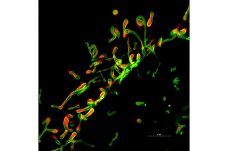
Cell with damaged mitochondria. Credit: ©Science China Press
Mitophagy plays a central role in the mitochondrial quality control system, and defective mitophagy is linked to a variety of human diseases. At present, how the damaged mitochondria are selectively recognized and removed to ensure the accuracy of mitophagic clearance remains unclear.
Miro2, a mitochondrial subfamily of the Ras GTPases, was shown to sense both the depolarization and the Ca2+ release from mitochondria, acting as a Parkin receptor for accurate removal of damaged mitochondria, according to a new study published by Science Bulletin.
PINK and Parkin are causal genes for autosomal recessive early-onset Parkinsonism and key components of mitochondrial quality control system. Parkin, an E3 ligase, translocates from cytoplasm to damaged mitochondria to ubiquitinate numerous mitochondrial outer membrane proteins, which is believed to be the tag for mitophagic clearance. Before translocating to mitochondria, Parkin is able to be activated by phosphorylated ubiquitin in addition to its phosphorylation by PINK1. Since ubiquitin phosphorylation by PINK1 takes place in the vicinity of the mitochondrial outer membrane, Parkin likely translocates proximal to the mitochondrial outer membrane in order to be activated by PINK1. Therefore, there could be mitochondrial outer membrane protein(s) supplying a platform for Parkin recognition and recruitment.
The authors propose a model on how Miro2 functions as a receptor for Parkin recognition and translocation to damaged mitochondria. Under unperturbed conditions, PINK1 is cleaved by PARL at mitochondrial inner membrane. Upon mitochondrial membrane potential collapse, Miro2 is quickly phosphorylated at Ser325/Ser430 by the full-length PINK1 after PINK1 translocates to the mitochondrial outer membrane. Meanwhile, Ca2+ efflux from the mitochondria is sensed by the EF2 hand domain of Miro2. When Miro2 is phosphorylated by PINK1 and binds with Ca2+ released from mitochondria, Miro2 undergoes a realignment and demultimerization from tetramers to monomers on the mitochondrial outer membrane. Then the realigned Miro2 acts as a platform for Parkin recognition, translocation, and subsequent initiation of mitophagy. Thus, Miro2 is able to detect both membrane potential collapse and Ca2+ release from damaged mitochondria to ensure that only damaged mitochondria are recognized and targeted by Parkin for mitophagic clearance.
Dysfunctional mitochondria undergo multiple changes such as depolarization, Ca2+ efflux, ROS elevation etc. Therefore, "it is important for mitochondrial Parkin receptors to be able to detect multiple signals from damaged mitochondria to ensure the accuracy of mitophagy", said Tie-Shan Tang, head of the research team.
In agreement with the crucial role of Miro2 in selective removal of damaged mitochondria, Miro2 defective mice showed delayed reticulocyte maturation, lactic acidosis, and cardiac disorders. "Cardiac dilatation was obviously seen in Miro2-KO hearts at 11-month-old, and the accumulation of abnormal mitochondria in cardiac myocytes was also significant in Miro2-KO mice", said Jiu-Qiang Wang, the first author of the paper.
"We provide biochemical, molecular and cellular, and in vivo mouse model data demonstrating that Miro2 functions as a platform for Parkin translocation and damaged mitochondria clearance. These findings shed new light on understanding the pathological mechanisms of human diseases such as neurodegenerative diseases and cardiac disorders", the authors concluded.
More information: Jiu-Qiang Wang et al, Miro2 supplies a platform for Parkin translocation to damaged mitochondria, Science Bulletin (2019). DOI: 10.1016/j.scib.2019.04.033
Provided by Science China Press


