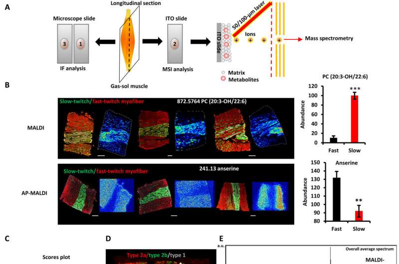
Reproducibility of metabolomic fingerprints of slow- and fast-twitch myofibers in MSI. (A) Schematic of the MSI workflow. Every three consecutive sections from the muscle were prepared as a group, with the first and third sections used for immunofluorescence (IF) analysis and the middle sections used for MSI analysis. (B) Fingerprint metabolites of slow- and fast-twitch myofibers detected by AP-MALDI-QTOF and MALDI-FTICR (left). Multicolor immunofluorescence staining of the GAS-SOL muscle (right). Means ± SEM; N = 3; **P < 0.01 and ***P < 0.001 compared with fast-twitch myofibers; Student’s t test. Green, slow twitch; red, fast twitch. Scale bars, 500 μm. (C) PLS-DA of the fast-twitch myofibers and the slow-twitch myofibers (N = 24 independent MSI experiments). (D) Performance of single-cell MSI on myofibers at the high spatial resolution of 25 μm. Immunofluorescence staining (top) and MSI analysis (bottom) of adjacent longitudinal sections of the hindlimb’s GAS-SOL muscle. Green, type 2a myofiber; red, type 2b myofiber; white, type 1 myofiber. Scale bar, 1000 μm. (E) Averaged mass spectrum from MALDI (top) and AP-MALDI (bottom) of the GAS-SOL muscle.ITO, indium tin oxide; a.u., arbitrary units. (F) Root mean square–normalized abundance and distributions of discriminative peaks (table S1) in slow- and fast-twitch myofibers according to MALDI- and AP-MALDI-MSI of GAS-SOL muscle (left). Fingerprint metabolites of slow- and fast-twitch myofibers detected by AP-MALDI-QTOF and MALDI-FTICR. Immunofluorescence staining and MSI of adjacent longitudinal sections of the hindlimb’s GAS-SOL muscle (right). Green, slow twitch; red, fast twitch. Credit: Science Advances (2023). DOI: 10.1126/sciadv.add0455
You might think that only DC Comics superhero The Flash could run at a speed of 200 strides per second. But in the animal world, special muscles—called "superfast muscles"—can move as fast as Barry Allen.
Now, for the first time, scientists from the Institute of Zoology and the Beijing Institute of Stem Cells and Regenerative Medicine of the Chinese Academy of Sciences have identified these muscles in mouse legs, raising the prospect of future research that could break the physical speed limits of normal human arm and leg movements.
Traditionally, superfast muscles have only been found in some obscure parts of animal anatomy including hummingbird wings, rattlesnake tails, and bat larynxes. While fast twitch muscles—the muscles that athletes use for activities like sprinting—are widespread in mammalian limbs, superfast muscles had previously only been identified in one part of human and mammalian anatomy: the extraocular muscles that control rapid eye movement.
"We achieved this feat by optimizing a new technology called single-cell metabolomic imaging," said Prof. Ng Shyh Chang, senior author of the study.
The researchers used this new technology to study cells in frozen slices of mouse leg muscles. They found that some of these cells contained metabolic signatures, i.e., groups of biochemicals produced during cell metabolism, that are normally found only in superfast muscles.
The superfast cells in the mouse legs, which showed a large amount of metabolites and genes associated with physical training, also contained metabolic signatures resembling those of oxidative muscles—i.e., fatigue-resistant muscles used for endurance exercise like marathons.
The scientists hypothesized that, with repeated neural stimulation, the mouse legs had begun to form small numbers of superfast muscles similar to those that control human eye movements.
With this knowledge, scientists may be able to unlock a wide range of therapeutic interventions that push the physical boundaries of what human limbs can achieve.
The research is published in the journal Science Advances.
More information: Lanfang Luo et al, Spatial metabolomics reveals skeletal myofiber subtypes, Science Advances (2023). DOI: 10.1126/sciadv.add0455
Journal information: Science Advances
Provided by Chinese Academy of Sciences


