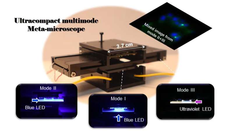
Photographic image of the multimode microscope. Credit: Advanced Devices & Instrumentation
Versatility and miniaturization of imaging systems are of great importance in today's information society. Microscopic imaging techniques have always been indispensable for scientific research and disease diagnosis in the biomedical field, which is also stepping towards the integration, portable, and multi-functions.
The optical microscope technique usually includes bright-field, dark-field, and fluorescence imaging, which are commonly based on cumbersome optical components. Especially for fluorescence and dark-field microscopy, blocking unwanted background stray light to ensure the high SNR is of significant importance in imaging performance.
The employment of guided-wave illumination makes it possible to combine two imaging techniques together, but the systems are still bulky and complicated. A promising route to a compact microscope is to use metalenses, which consist of subwavelength nanostructures with powerful capabilities to modulate the amplitude and phase of light.
Although innovative metalenses have been demonstrated for fluorescence microscopy, the advantages of ultrathin and flat architecture have not been revealed yet for miniaturization.
Professor Tao Li & Shining Zhu's group from Nanjing University reported a miniaturized multimode microscope for bright-field, dark-field, and fluorescence imaging by introducing guided-wave illuminations. By conveniently switching the light source, three imaging modes can work together or separately within a very compact microscope (several centimeters in size).
Notably, the proposed guided-wave illumination module not only provides a low noise imaging mode, but also further reduces the system size that favors the compact microscope very much.
As a result, a metalens array is designed and fabricated with a magnification of 3.5× in imaging (working at λ = 470 nm), which corresponds to the emission wavelength of fluorescence imaging. The imaging resolution is approximately 714 nm, ensuring subcellular imaging. Moreover, experiments have demonstrated the potential applications of microfluidic imaging techniques to miniaturize microfluidic imaging systems further.
In conclusion, the researchers propose and demonstrate a miniaturized multimode meta-microscope based on guided-wave illumination. Three imaging modes are realized within a centimeter-scale prototype, including bright-field, dark-field, and fluorescence modes.
The proposed guided-wave illumination further saves the space to meet this compactness, which significantly combines dark-field and fluorescence imaging together. A metalens array is particularly designed and works in a zoom-in mode (3.5×) incorporated with a CMOS image sensor, which is designed with respect to the wavelength of 470 nm corresponding to the emission wavelength.
The half-pitch resolution is about 714 nm, ensuring sub-cellular imaging resolution. Notably, this is the first meta-device implementation of multimode imaging in an ultracompact system, which is expected to enable real-time visualization of cell culture and have a great impact in the biomedical field in the future.
The paper is published in the journal Advanced Devices & Instrumentation.
More information: Xin Ye et al, Ultracompact Multimode Meta-Microscope Based on Both Spatial and Guided-Wave Illumination, Advanced Devices & Instrumentation (2023). DOI: 10.34133/adi.0023
Provided by Advanced Devices & Instrumentation


