New sensor for SERS Raman spectroscopy almost as sensitive as a dog's nose

Using carbon nanotubes, a research team led by Professor Hyung Gyu Park in collaboration with Dr. Tiziana Bond has developed a sensor that greatly amplifies the sensitivity of commonly used but typically weak vibrational spectroscopic methods, such as Raman spectroscopy. This type of sensor makes it possible to detect molecules present in the tiniest of concentrations.
Scientists at ETH Zurich and the Lawrence Livermore National Laboratory (LLNL) in California have developed an innovative sensor for surface-enhanced Raman spectroscopy (SERS). Thanks to its unique surface properties at nanoscale, the method can be used to perform analyses that are more reliable, sensitive and cost-effective. In experiments with the new sensor, the researchers were able to detect a certain organic species (1,2bis(4-pyridyl)ethylene, or BPE) in a concentration of a few hundred femtomoles per litre. A 100 femtomolar solution contains around 60 million molecules per liter.
Until now, the detection limit of common SERS systems was in the nanomolar range, i.e. one billionth of a mole. The results of a study conducted by Hyung Gyu Park, Professor of Energy Technology at ETH Zurich, and Tiziana Bond, Capability Leader at LLNL, were published this week as a cover article in the scientific journal Advanced Materials.
Raman spectroscopy takes advantage of the fact that molecules illuminated by fixed-frequency light exhibit 'inelastic' scattering closely related to the vibrational and rotational modes excited in the molecules. Raman scattered light differs from common Rayleigh scattered light in that it has different frequencies than that of the irradiating light and produces a specific frequency pattern for each substance examined, making it possible to use this spectrum information as a fingerprint for detecting and identifying specific substances. To analyse individual molecules, the frequency signals must be amplified, which requires that the molecule in question either be present in a high concentration or located close to a metallic surface that amplifies the signal. Hence the name of the method: surface-enhanced Raman spectroscopy.
Amplified signals for improved reproducibility
"This technology has been around for decades," explains Ali Altun, a doctoral student in the group led by Park at the Institute of Energy Technology. With today's SERS sensors, however, the signal strength is adequate only in isolated cases and yields results with low reproducibility. Altun, Bond and Park therefore set themselves the goal of developing a sensor that massively amplifies the signals of the Raman-scattered light.
The substrate of choice turned out to be vertically arranged, caespitose, densely packed carbon nanotubes (CNT) that guarantee this high density of 'hot spots'. The group developed techniques to grow dense forests of CNTs in a uniform and controlled manner. The availability of this expertise was one of the principal motivations for using nanotubes as the basis for highly sensitive SERS sensors, says Park.

A spaghetti-like surface
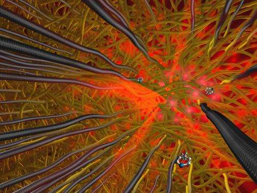
The tips of the CNTs are sharply curved, and the researchers coated these tips with gold and hafnium dioxide, a dielectric insulating material. The point of contact between the surface of the sensor and the sample thus resembles a plate of spaghetti topped with sauce. However, between the strands of spaghetti, there are numerous randomly arranged holes that let through scattered light, and the many points of contact—the 'hot spots'—amplify the signals.
"One method of making highly sensitive SERS sensors is to take advantage of the contact points of metal nanowires," explains Park. The nano-spaghetti structure with metal-coated CNT tips is perfect for maximising the density of these contact points.
Indeed, Bond explains, the wide distribution of metallic nano-crevices in the nanometre range, well recognised to be responsible for extreme electromagnetic enhancement (or hot spots) and highly pursued by many research groups, has been easily and readily achieved by the team, resulting in the intense and reproducible enhancements.
The sensor differs from other comparable ultra-sensitive SERS sensors not only in terms of its structure, but also because of its relatively inexpensive and simple production process and the very large surface area of the 3D structures producing an intense, uniform signal.
A breakthrough on two levels
Initially, the researchers only coated the tips of the CNTs with gold. The first experiments with the BPE test molecule showed them that they were on the right track, but that the detection limit could not be reduced to quite the degree they had hoped. Eventually, they discovered that the electrons required on the gold layer surface for generating what is referred to as plasmon resonance were flowing out via the conductive carbon nanotubes. The task was then to figure out how to prevent this plasmonic energy leakage.
The researchers coated the CNTs with hafnium oxide, an insulating material, before applying a layer of gold. "This was the breakthrough," says Altun. The insulation layer increased the sensitivity of its sensor substrate by a factor of 100,000 in the molar concentration unit.
"For us as scientists, this was a moment of triumph," agrees Park, "and it showed us that we had made the right hypothesis and a rational design."
The key to the successful development of the sensor was therefore twofold: on the one hand, it was their decision to continue using CNTs, whose morphology is essential for maximising the number of 'hot spots', and on the other hand, it was the fact that these nanotubes were double-coated.
Park and Bond would now like to go one step further and bring their new principle to market, but they are still seeking an industry partner. Next, they want to continue improving the sensitivity of the sensor, and they are also looking for potential areas of application. Park envisions installation of the technology in portable devices, for example to facilitate on-site analysis of chemical impurities such as environmental pollutants or pharmaceutical residues in water. He stresses that invention of a new device is not necessary; it is simple to install the sensor in a suitable way.
Other potential applications include forensic investigations or military applications for early detection of chemical or biological weapons, biomedical application for real-time point-of-care monitoring of physiological levels, and fast screening of drugs and toxins in the area of law enforcement.
More information: Altun AO, Youn SK, Yazdani N, Bond T, Park HG. 'Metal-Dielectric-CNT Nanowires for Femtomolar Chemical Detection by Surface Enhanced Raman Spectroscopy.' Advanced Materials, 2013. DOI: 10.1002/adma.201300571
Journal information: Advanced Materials
Provided by ETH Zurich


