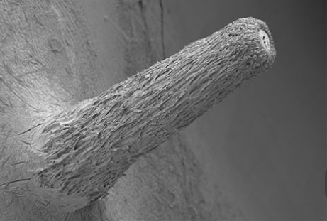
The images were obtained using cryo scanning electron microscopy, where the sample is plunged into liquid nitrogen to freeze it and imaged using the electron microscope.
The benefit of this method is that the sample is imaged in as close to its natural state as possible, providing the best quality 3D view of an organism.
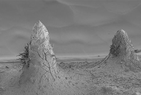
We want to understand how the fungus makes the fruiting bodies (toadstools) that produce the spores called ascospores which cause infection of ash. The fungus growing within the ash leaf stem produces sex organs and that the images show different stages of growth of such an organ. We think that this organ may be fertilised to initiate the sexual cycle that produces infective ascospores.
Provided by John Innes Centre


