This article has been reviewed according to Science X's editorial process and policies. Editors have highlighted the following attributes while ensuring the content's credibility:
fact-checked
peer-reviewed publication
trusted source
proofread
New tool to detect protein-protein interactions could lead to promising avenues for gene therapy and other treatments

SMU nanotechnology expert MinJun Kim and his team have developed a faster, more precise way to detect the properties and interactions of individual proteins crucial in rapid, accurate, and real-time monitoring of virus-cell interactions. This could pave the way for innovative medical therapies and advancements to be created using gene therapy—a technique that utilizes harmless viruses to modify a person's genes to treat or cure disease.
Beyond that, this research could also be used to detect and characterize other types of protein-protein interactions, potentially leading to the development of treatments that can regulate interactions causing adverse effects in the body, said Kim, the Robert C. Womack Chair in the Lyle School of Engineering at SMU and principal investigator of the BAST Lab.
A study published in the journal Nanoscale shows that this tiny device Kim's team created accurately determines in real-time when two proteins that play a role in targeted gene therapy—known as fibroblast growth factor (FGF-1) and heparin—have bonded with each other.
And unlike the ways protein-protein interactions are detected now, this device only needs a small sample size to investigate the properties of individual proteins and their complex interactions, saving time and cost for the analysis.
Proteins are the workhorses that facilitate most biological processes in a cell. Often, it's necessary for two or more proteins to bind with each other—meaning they've connected with each other as a result of biochemical events—to carry out certain functions.
That's the case with proteins FGF-1 and heparin.
Together, these proteins have been shown to help a harmless virus called adeno-associated viruses (AAV)—which is the go-to vehicle for gene therapy—latch on to the right cell receptors in the human body.
Viral gene therapy uses viruses like AAVs as a way to deliver a healthy copy of a gene into a person to replace or modify a disease-causing one. But the problem is that AAVs have several different types, or serotypes, and each one has a natural preference to infect and thrive in specific tissue types, such as those serving the heart or kidneys. That means that for gene therapy to be successful in unloading the virus' cargo to its intended target, the right serotype of AAV needs to bond with the correct cell receptors.
Yet, not enough is currently known about how this process called tropism works to ensure that.
"Thus, a better understanding of heparin and FGF-1 interactions will help us comprehend tropism for AAV gene therapy," which, in turn, could make it possible to target new gene therapies for specific diseases, Kim said.
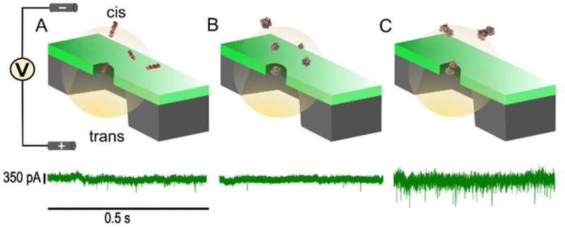
Kim's team created and tested a device known as a solid-state nanopore, which can accurately tell when heparin and FGF-1 have bonded.
How the device works
Nanoparticles are too small to be visible to the naked eye—ranging in size from 1 to 100 nanometers (one billionth of a meter) in size. Nanomaterials can occur naturally and can also be engineered to perform specific functions, such as the delivery of drugs to various forms of cancer.
Each nanopore in this study was made from 12-nanometer-thick silicon nitride (SixNy) membranes, with a hole of roughly 17 nanometers in diameter drilled through it.
These so-called solid-state nanopores were able to tell when heparin bonded with FGF-1, because Kim and his team have calculated the electrical currents of three different scenarios: when only heparin is present in the sample; when only FGF-1 is present; and when there is an equal ratio of the two proteins.
How does the device know what the electrical current is?
Basically, a molecule from the sample passes through a tiny hole in the device that separates two chambers containing electrolyte solutions. This leads to fluctuations in the electrical current, which can be decoded to detect heparin-FGF-1 bonding.
Kim said, "the findings of this research represent a preliminary experiment laying the groundwork for future endeavors."
His ultimate goal is to be able to use solid-state nanopores on two other proteins also known to be important for targeted gene therapy: the actual binding of the AAVs with cell surface receptors.
AAVs have a protein coat called a capsid that surrounds their genetic information, which is what gets altered by gene therapists to introduce a new healthy gene into a person. It is only when capsids bind with cell receptors—another protein found on the surface of cells—that the virus and cell are connected and the virus' cargo can be released.
"The effectiveness of targeted gene therapy depends on the affinity between virus capsid and cell surface receptors," Kim explained.
Kim wants to be able to use solid-state nanopores to measure that, making it clearer when a virus has successfully delivered its cargo into a person. That's because a key barrier to using viral gene therapy is that the amount of genetic material transmitted by AAV can't be measured, potentially leading to overdosing or underdosing.
In addition to making breakthroughs in gene therapy, lead study author Navod Thyashan, a graduate research assistant at SMU's BAST, noted that these nanopores could also set the stage for other new medical treatments to be developed. It can be used with other proteins known to have a high affinity for bonding with each other, allowing for treatments to potentially regulate these interactions that cause diseases.
"Solid-state nanopores (SSNs) can be fabricated in sizes ranging from single digit nanometers in diameter to hundreds," he said. "Thus, SSNs can be used in most biomolecule sensing applications, as long as we choose the correct nanopore diameter for the proteins we are dealing with."
Helping Thyashan and Kim create the device were Madhav L. Ghimire, the Dean's Postdoctoral Fellow at SMU's Moody School of Graduate and Advanced Studies; and Sangyoup Lee, with the Bionic Research Center for the Korea Institute of Science and Technology.
More information: Navod Thyashan et al, Exploring single-molecule interactions: heparin and FGF-1 proteins through solid-state nanopores, Nanoscale (2024). DOI: 10.1039/D4NR00274A
Journal information: Nanoscale
Provided by Southern Methodist University


