This article has been reviewed according to Science X's editorial process and policies. Editors have highlighted the following attributes while ensuring the content's credibility:
fact-checked
peer-reviewed publication
trusted source
proofread
Method can analyze individual, still-living cells that may contain biomarkers for cancer and other deadly diseases

The Ivanov Lab at Northeastern University is paving the way to a whole variety of diagnostic tests that are possible off of a single blood draw, including—someday—cancer.
Every cell in your body has a thin covering of carbohydrates called glycoconjugates—"conjugates" as they're formed when the carbohydrates are chemically connected to proteins, lipids and other cell surface molecules.
These carbohydrates—also called glycans—play a significant role in both cell communication and the cell's ability to respond to disease.
For instance, says associate professor of chemistry and chemical biology Alexander Ivanov, common blood type tests "are based on glycan analysis."
But that's far from the only thing glycans can show us. "We see that the glycans reflect cell types and cell states," he continues, which are "potentially diagnostic of different diseases."
"Cancer is definitely among those."
Abnormalities in the presentation of glycans on the cell surface, their types and composition, their chemical linkages and their quantities, all provide potentially useful information to the researcher or diagnostician, including biomarkers for a variety of diseases.
Now, a team of researchers led by Ivanov has developed a method by which they can analyze the surface glycans of an individual, still-living human cell, as well as those found in minuscule volumes of plasma and other blood isolates.
Biomarkers and new technologies
Within the glycome—the overall representation of glycans in a cellular environment, especially on the cell surface—"abnormalities represent an overt source of potential biomarkers for the diagnostic, prognostic, and treatment monitoring of various human diseases," they write in a paper published in Nature Communications.
"We're working on new technologies to enable high-sensitivity analysis of proteome systems," which refers to how proteins function in the body, Ivanov says. "Native proteomics, top-down proteomics, bottom-up proteomics.
"Some years ago, we also began to look at other biological molecules, in addition to proteins."
"The main theme of our research," he continues, "is developing new sample preparation and separation technologies coupled to mass spectrometry to analyze complex biological samples."
"In the human organism, we have above 20,000 genes," Ivanov says, many of which encode proteins that "are responsible for very diverse, multiple biological functions," enabled through chemical changes called "post-translational modifications."
"The protein, which corresponds to a gene or gene product, gets modified," Ivanov explains, "and through different post-translational modifications the properties, activity and functions of those proteins can change dramatically. And glycosylation," whereby a protein is modified by a glycan, "is one of the most common and, arguably, the most prominent post-translational modification."
"This is why analyzing those glycan functionalities attached to proteins—as well as other molecules—can be very informative for understanding the biology of the cell in health and disease."
The Ivanov Lab's new separation tools and analytical protocols can help isolate the wide diversity of glycans involved in cellular functions and potentially identify biomarkers that might indicate whether the cell has been altered by diseases like cancer, autoimmune disorders or neurodegenerative conditions like Alzheimer's.

Experiments in capillaries
One of the principal interventions in their study, says Anne-Lise Marie, an associate research scientist in the Ivanov Lab, is their ability to observe cells that are still alive. With this method, "we preserve the native state of the biological samples and their biomolecular constituents," she says, which means the researchers can "analyze glycans in their native, intact form," within their original environment.
Previous methods for characterizing glycans "relied on various chemical derivatization techniques," Ivanov wrote in a follow up, which led to a loss of contextual data, "suboptimal labeling performance, glycan degradation, sample loss and inaccurate quantitation."
"Every day we discover new types of glycans," Marie says, including those of "extremely low abundance."
In another study, published in Nature Communications last year, the Ivanov Lab detailed a new, highly sensitive technique that makes the analysis of native glycans possible.
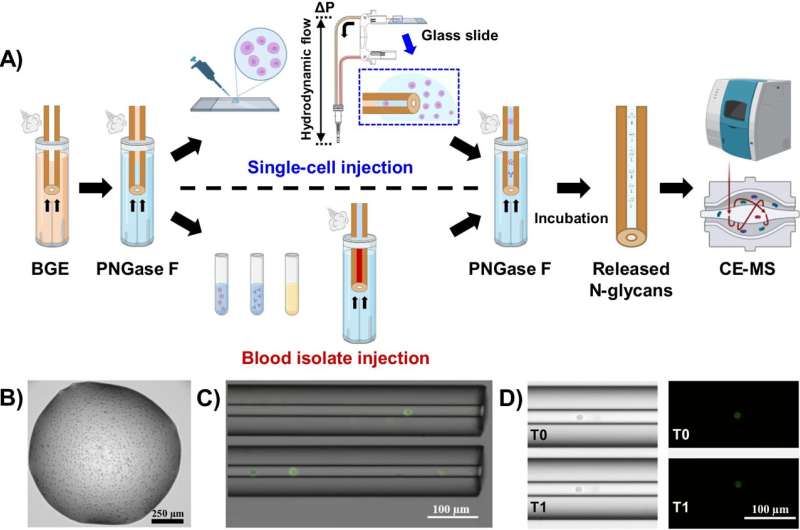
"Everything is prepared in capillaries," says Ph.D. candidate Yunfan Gao. Using very thin glass straws—with apertures less than half the diameter of a human hair—Gao isolates an individual cell from the rest of the sample, drawing it into this "capillary."
The capillary is then used to release and separate the glycans before analysis by a mass spectrometer.
"The separation method, called 'capillary electrophoresis,' pulls apart the components [of the cell sample], based on the differences in their charge and size," Ivanov wrote.
When the team began experimenting with in-capillary sample preparation, they didn't know if the technique would be "sensitive enough to analyze such a small amount from a single cell," Gao says.
"No one has directly analyzed, quantified and structurally characterized intact proteins and native glycans from a single cell."
"In-capillary techniques," he continues, are the best "way that we can minimize sample loss."
"I am pretty convinced that the in-capillary option is the best way to go," Marie agrees. When they first moved to this method, she says, "I was happy to see that we detected [a large variety] of glycans released from just one single cell."
The process results in "crazy sensitivity," she says.
What comes next
Nevertheless, there are limits to what single-cell analysis can accomplish. Gao notes that there is inherently a "limited amount of the total cellular contents" of an individual cell—one cell can only store so much information.
The lab's next direction is to "target multiple modalities of biological molecules in single cells," not just glycomes, Gao continues. That way, "we can make full use of the whole cell [and] maybe get a better and more complete picture of the cell state."
That, Ivanov says, and automating the process of cell isolation and separation. "This will bring us closer to enabling new biological studies and clinical applications, which is super exciting."
In direct single-cell analysis of glycomes and intact proteomes, Ivanov says, "we have published first in these two areas," and now the race is on. "We opened these areas for active research, which other labs are trying to pursue now."
More information: Anne-Lise Marie et al, Native N-glycome profiling of single cells and ng-level blood isolates using label-free capillary electrophoresis-mass spectrometry, Nature Communications (2024). DOI: 10.1038/s41467-024-47772-w
Journal information: Nature Communications
Provided by Northeastern University
This story is republished courtesy of Northeastern Global News news.northeastern.edu.


