Cellular memory of stressful situations

Stress is unhealthy. The cells use therefore a variety of mechanisms to deal with stress and avert its immediate threat. However, certain stressful situations leave marks that go beyond the immediate response; some even seem to be passed on to the next generation. One school of thought that has gained a lot of attention lately, hypothesizes that the epigenome, chemical modifications on the DNA and on proteins, carries information about external influences such as stress. However, when and how environmental cues trigger changes in the epigenome and thus influence our response has remained largely unknown.
To investigate how the environment can influence epigenetic processes, FMI Senior group leader Marc Bühler and his group work with fission yeast. In a paper published in Cell Reports, they report the identification of a regulatory circuit that buffers a potentially deleterious epigenetic switch under stressful conditions. The capacity to prevent such an epigenetic change is important to the organism, perhaps because the altered epigenetic state is maintained for many cell divisions, well after the stress has subsided.
Unicellular organisms such as fission yeast can be easily stressed with high temperatures, which are commonly encountered in the wild. One highly conserved protein that is particularly sensitive to high temperatures is Dicer. Under normal circumstances, it sits in the nucleus and—among other things—inhibits the expression of genes that deal with stress responses. If fission yeast is exposed to high temperatures, Dicer leaves the nucleus and accumulates in the cytoplasm. This allows expression of the stress response genes. Thus, by moving out of the nucleus, Dicer enables full activation of the yeast's stress response.
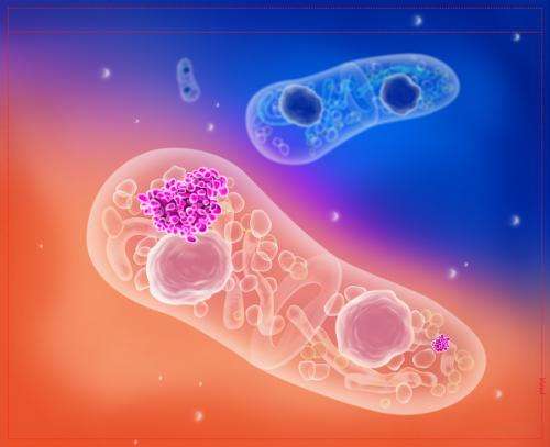
In their study, Bühler and colleagues used correlative light and electron microscopy (CLEM) to show that Dicer accumulates as an aggregate in a novel cytoplasmic compartment during heat stress. There, Dicer is joined by the well-known protein disaggregase Hsp104, which dissolves the Dicer aggregates. Dicer then recycles back to the nucleus. "What is particularly intriguing is that Hsp104 is one of the "stress genes" that is repressed by Dicer in the nucleus. Hsp104 thus guarantees that Dicer can again take on its nuclear functions, one of them being the repression of "stress genes" like HSP104. Thus we are dealing a with classical negative feedback loop", explains Bühler.
Which brings us to the other highly relevant function of Dicer in this context: Dicer ensures the stability of the yeast genome by maintaining the dense packaging of the centromeric regions into heterochromatin. Without heterochromatin, chromosomes segregate improperly during mitosis. This can result in aneuploidy and chromosome instability, which are a hallmark of cancer. In their paper, the FMI scientists show that in the absence of Hsp104, Dicer remains in the cytoplasm and heterochromatic regions on the yeast genome are no longer stable after exposure to heat stress. This leads to changes in gene expression and most deleteriously, chromosome missegregation during cell division. "The observed changes in heterochromatin and gene expression incurred after a stressful situation in the absence of functional Hsp104, are still present long after the stress has subsided," comments Bühler. "What we have here is a phenomenon that complies with the classic definition of epigenetics: an event where an environmental cue triggers a change in gene expression that does not entail a change in the genomic sequence, yet which is inherited for many cell divisions, even after the environmental cue has disappeared."
In an interesting twist, Bühler and his team also showed that the feedback loop that controls Dicer and Hsp104, plays a role during prion formation and propagation. In other organisms, Hsp104 was well-knownfor its function in the aggregation of prionogenic proteins. Indeed, the FMI scientists found that without Dicer, Hsp104 is off its leash and increases the frequency of toxic prionogenic protein aggregates that eventually lead to cell death. "Given that protein aggregates play an important role not only in prion diseases but also in neurodegenerative disorders, the processes described in yeast may become an interesting avenue for future studies into diseases like Alzheimers," comments Bühler.
At the frontier of microscopy: Correlative Light and Electron Microscopy (CLEM) & Serial Block Face Scanning Electron Microscopy (SBFEM)
- SBFEM allows the three dimensional reconstruction of the structures within a cell and tissues at a high resolution (see picture of the cell). An ultramicrotome integrated within the specimen chamber of a scanning electron microscope cuts away a thin slice of the specimen, only 50-100 nanometers thick, thus allowing the instrument to scan the next "layer". Sequential 2D images are then assembled to a 3D image of the cell and its structures. For their project, the scientists profited substantially from in-house expertise at the Facility for Advanced Imaging and Microscopy (FAIM), namely Christel Genoud, who is a leading expert in SBFEM.
- CLEM is a highly sophisticated microscopy method that is mastered by only few laboratories around the world. It combines fluorescent light microscopy with electron microscopy, two platforms that are normally separated. It thus takes advantage of both technologies: light microscopy allows the study of processes in whole, often living, cells for example with fluorescent labels. Electron microscopy shows ultrastructural detail in parts of cells at a very high resolution. CLEM combines the advantages of both thus adding detail and providing complementary information. An ERC Starting Grant made the introduction of CLEM at the FMI possible.
More information: Oberti D, Biasini A, Kirschmann MA, Genoud C, Stunnenberg R, Shimada Y, Bühler M (2014) "Dicer and hsp104 function in a negative feedback loop to confer robustness to environmental stress." Cell Rep. 2015 Jan 6;10(1):47-61. DOI: 10.1016/j.celrep.2014.12.006. Epub 2014 Dec 24.
Journal information: Cell Reports


