This article has been reviewed according to Science X's editorial process and policies. Editors have highlighted the following attributes while ensuring the content's credibility:
fact-checked
peer-reviewed publication
trusted source
proofread
Correlative light electron microscopy using gold nanoparticles as single probes

Correlative light electron microscopy (CLEM) is a powerful tool in bioimaging, as it combines the ability to image living cells over large fields of view with molecular specificity using light microscopy (LM) with the high spatial resolution and ultrastructural information of electron microscopy (EM). To highlight biomolecules of interest and determine their position with high accuracy in CLEM, researchers must ensure they are labeled with probes that are visible both in LM (typically by fluorescence) and in EM (using electron dense material). However, existing probes have a number of drawbacks, including lack of stability under LM (photobleaching) and lack of integrity.
In a new paper published in Light Science & Application, a team of scientists, led by Professor Paola Borri from Cardiff University and Professor Paul Verkade from the University of Bristol, United Kingdom, has developed a new CLEM approach that uses small gold nanoparticles as a single probe visible in both LM and EM with high contrast and photostability, with no need to use fluorescent probes.
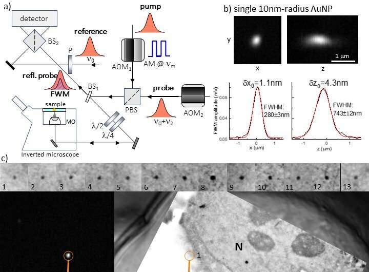
For LM, they exploited an optical microscope developed in Professor Borri's group. This microscope detects the non-linear optical response of gold nanoparticles, called four-wave mixing (FWM). With this method, the researchers were able to detect individual small gold nanoparticles as single probes of the epidermal growth factor protein in human cancer cells, located with nanometric precision background-free by light microscopy, and correlatively mapped with high accuracy to the corresponding transmission electron microscopy image.
Owing to the background-free and photostable FWM response of individual gold nanoparticles, well visible in EM due to their electron dense composition, FWM-CLEM opens new avenues for highly accurate correlative microscopy workflows without the need to use unstable fluorophores or additional fiducial markers, thus greatly simplifying sample preparation protocols.
The scientists summarize the operational principle of their FWM microscope as follows: "In its general form, FWM is a third-order non-linear light-matter interaction phenomenon wherein three light fields interact in a medium to generate a fourth wave. Here, we use a scheme where all light waves have the same center frequency, in resonance with the optical absorption peak of a gold nanoparticle. For excitation and detection, we use a combination of short optical pulses of about 150fs duration, called pump and probe, generated by the same laser source. The detected FWM can be understood as a pump-induced change in the ability of the gold nanoparticle to transmit and scatter light, which manifests as a change in the probe beam scattered by the particle in the presence of the pump.
"FWM detection is remarkably photo-stable and high contrast. With this method we are able to detect individual small (down to 5nm radius) gold nanoparticles inside scattering and auto fluorescing cells and tissues completely free from background, at imaging speeds and excitation powers compatible with live cell imaging, with a sensitivity limited only by photon shot noise. We have also seen that FWM is a sensitive reporter of gold nanoparticle shapes, promising for multiplexing by shape recognition in future applications."
The researchers conclude, "FWM-CLEM opens new exciting possibilities for correlative light electron microscopy workflows, overcoming existing limitations with fluorescent probes and/or fiducial markers which are not needed in our method. Generally, we believe that FWM-CLEM will have a widespread impact. For example, combined with existing strategies to label with or even encapsulate gold nanoparticles inside virions, FWM opens the exciting prospect to track single virions over long observation times, background-free and deep inside living cells and tissues, to then pin-point events of interest (e.g. genome release) in the context of the cellular ultrastructure by CLEM."
More information: Iestyn Pope et al, Correlative light-electron microscopy using small gold nanoparticles as single probes, Light: Science & Applications (2023). DOI: 10.1038/s41377-023-01115-4
Journal information: Light: Science & Applications
Provided by Chinese Academy of Sciences


