Slow-moving shell of water can make Parkinson's proteins 'stickier'

Water—which makes up the majority of every cell in the body—plays a key role in how proteins, including those associated with Parkinson's disease, fold, misfold, or clump together, according to a new study.
When attempting to discover potential treatments for protein misfolding diseases, researchers have primarily focused on the structure of the proteins themselves. However, researchers led by the University of Cambridge have shown that a thin shell of water is key to whether a protein begins to clump together, or aggregate, forming the toxic clusters which eventually kill brain cells.
Using a technique known as Terahertz spectroscopy, the researchers have shown that the movement of the water-based shell surrounding a protein can determine whether that protein aggregates or not. When the shell moves slowly, proteins are more likely to aggregate, and when the shell moves quickly, proteins are less likely to aggregate. The rate of movement of the shell is altered in the presence of certain ions, such as salt molecules, which are commonly used in the buffer solutions used to test new drug candidates.
The significance of the water shell, known as the hydration or solvation shell, in the folding and function of proteins has been strongly disputed in the past. This is the first time the solvation shell has been shown to play a key role in protein misfolding and aggregation, which could have profound implications in the search for treatments. The results are reported in Angewandte Chemie International Edition.
When developing potential treatments for protein misfolding diseases such as Parkinson's and Alzheimer's disease, researchers have been studying compounds which can prevent the aggregation of key proteins: alpha-synuclein for Parkinson's disease or amyloid-beta for Alzheimer's disease. To date however, there are no effective treatments for either condition, which affect millions worldwide.
"It's the amino acids that determine the final structure of a protein, but when it comes to aggregation, the role of the solvation shell, which sits on the outside of a protein, has been overlooked until now," said Professor Gabriele Kaminski Schierle from Cambridge's Department of Chemical Engineering and Biotechnology, who led the research. "We wanted to know whether this water shell plays a role in protein behavior—it's been a question in the field for a while, but no one has been able to prove it."
The solvation shell slides around on the surface of the protein, acting like a lubricant. "We wondered whether, if the movement of water molecules was slower in the solvation shell of a protein, it could slow the movement of the protein itself," said Dr. Amberley Stephens, the paper's first author.
To test the role of the solvation shell in the aggregation of proteins, the researchers used alpha-synuclein, the key protein implicated in Parkinson's disease. Using Teraheartz spectroscopy, a powerful technique to study the behavior of water molecules, they were able to observe the movement of the water molecules that surround the alpha-synuclein protein.
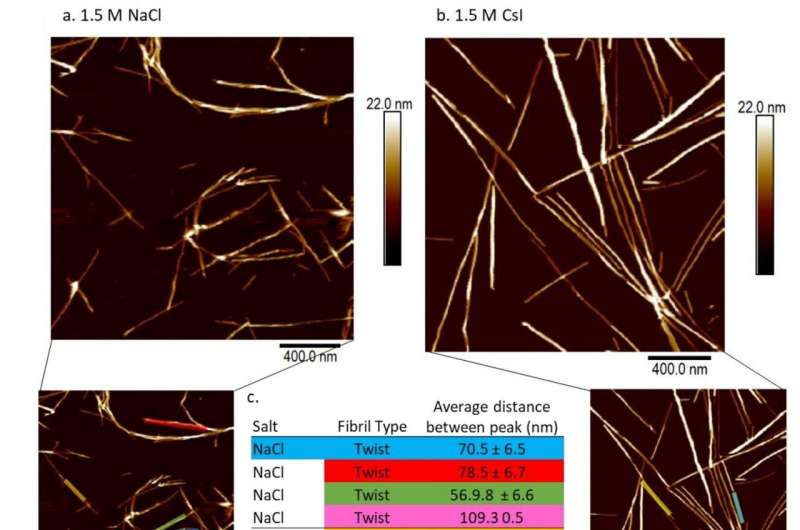
They then added two different salts in solution to the proteins: sodium chloride (NaCl), or regular table salt, and cesium iodide (CsI). The ions in the sodium chloride—Na+ and Cl-—bind strongly to the hydrogen and oxygen ions in water, while the ions in the cesium iodide make much weaker bonds.
The researchers found that when the sodium chloride was added, the strong hydrogen bonds caused the movement of the water molecules in the solvation shell to slow down. This resulted in slower movement of the alpha-synuclein, and the aggregation rate increased. Conversely, when the cesium iodide was added, the water molecules sped up, and the aggregation rate decreased.
"In essence, when the water shell slows down, the proteins have more time to interact with each other, so they're more likely to aggregate," said Kaminski Schierle. "And on the flip side, when the solvation shell moves more quickly, the proteins become harder to catch, so they're less likely to aggregate."
"When researchers are screening for an aggregation inhibitor for Parkinson's disease, they will usually use a buffer composition, but there's been very little thought on how that buffer is interacting with the protein itself," said Stephens. "Our results show that you need to understand the composition of the solvent inside the cell in order to mimic the conditions you have in the brain and ultimately end up with an inhibitor that works."
"It's so important to look at the whole picture, and that hasn't been happening," said Kaminski Schierle. "To effectively test whether a drug candidate will work in a patient, you need to mimic cellular conditions, which means you need to take everything into consideration, like salts and pH levels. The failure to look at the whole cellular environment has been limiting the field, which may be why we haven't yet got an effective treatment for Parkinson's disease."
More information: Amberley D. Stephens et al, Decreased Water Mobility Contributes To Increased α‐Synuclein Aggregation, Angewandte Chemie International Edition (2022). DOI: 10.1002/anie.202212063
Journal information: Angewandte Chemie International Edition
Provided by University of Cambridge


