This article has been reviewed according to Science X's editorial process and policies. Editors have highlighted the following attributes while ensuring the content's credibility:
fact-checked
peer-reviewed publication
trusted source
proofread
Electrical impedance tomography plus extracellular voltage activation technique simplifies drug screening

When developing new drugs, understanding their effects on ion channels in the body, such as the human ether-a-go-go-related gene (hERG) ion channel found in neurons and heart muscle cells, is critical. Blocking hERG channels can disrupt a normal heart rhythm, potentially leading to a fatal condition known as torsade de pointes.
Current methods for assessing these effects typically involve invasive procedures like patch-clamp techniques or fluorescence microscopy. These methods alter cell properties and may affect measurement accuracy, requiring specialized equipment and expertise, which increases cost and complexity.
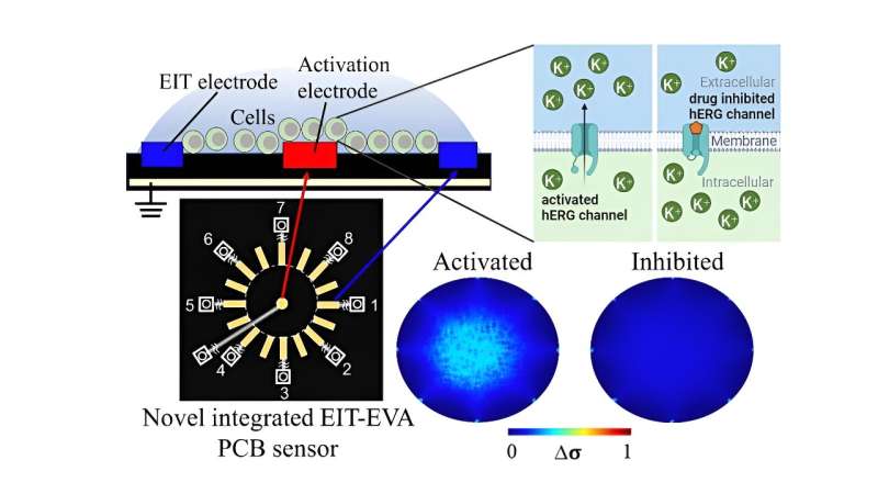
To address these challenges, researchers led by Daisuke Kawashima, an Assistant Professor at the Graduate School of Engineering at Chiba University, have proposed a novel, non-invasive method for real-time evaluation of drug effects on hERG channels. The work is published in the journal Lab on a Chip.
They developed a printed circuit board (PCB) sensor integrating electrical impedance tomography (EIT) with extracellular voltage activation (EVA). EIT measures impedance changes caused by ion movement, offering spatial information about extracellular ion distribution. EVA involves applying controlled extracellular voltages to induce changes in ion channel activity.
This integrated approach allows researchers to non-invasively activate hERG channels and monitor real-time ion flow changes in response to drug exposure.
More information: Muhammad Fathul Ihsan et al, Non-invasive hERG channel screening based on electrical impedance tomography and extracellular voltage activation (EIT–EVA), Lab on a Chip (2024). DOI: 10.1039/D4LC00230J
Journal information: Lab on a Chip
Provided by Chiba University


