March 18, 2024 report
This article has been reviewed according to Science X's editorial process and policies. Editors have highlighted the following attributes while ensuring the content's credibility:
fact-checked
peer-reviewed publication
trusted source
proofread
Using a non-destructive, light diffraction fingerprint technique to detect viral infections in cells

A combined team of engineers from Jiangsu University and Harvard University used a non-destructive, light diffraction fingerprint technique to detect viral infections in cells. Their paper is published in the journal Science Advances.
Currently, detecting viral infections in livestock involves the use of in vitro assays, applying chemicals to cells and studying the results under a microscope. Such work is typically expensive and time consuming, sometimes taking as long as 40 hours. The medical community has been asking the research community to find a better way.
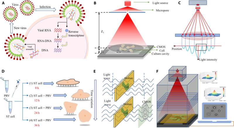
In this new study, the research team observed that when a virus infects a cell, tissue stress typically results. This led them to consider using lensless light diffraction to detect diffraction patterns for infected cells, an approach that would allow them to test cells quickly and without causing any damage.
Light from a lamp was shone through a micropore onto a slide holding a tissue sample. Beneath the cell was a CMOS sensor for collecting diffracted light. Data from the sensor was then sent to a computer for processing. Software isolated contrast and differential movement to create a diffraction fingerprint for a given cell in the tissue sample, which could then be compared to known fingerprints of cells infected with viruses.
The technique allows not only the identification of a virus by its diffraction fingerprint, but also the continuous monitoring of the same samples to determine damage extent.
The approach was highly accurate, with success rates roughly equivalent to those obtained by current standard testing methods. The researchers noted that analyzing a single sample typically took two hours or less and that it was nondestructive. It also could be automated. And because the test is based on software analysis, testing can be performed by nonprofessionals.
The researchers suggest that with further refinement, it could be used on site to test livestock or, looking forward, in test labs dedicated to detecting viruses in humans. These findings could prove critically important when the next pandemic strikes.
More information: Tongge Li et al, Virus detection light diffraction fingerprints for biological applications, Science Advances (2024). DOI: 10.1126/sciadv.adl3466
Journal information: Science Advances
© 2024 Science X Network


