This article has been reviewed according to Science X's editorial process and policies. Editors have highlighted the following attributes while ensuring the content's credibility:
fact-checked
peer-reviewed publication
trusted source
proofread
Q&A: Unveiling a new era of imaging—engineers lead breakthrough microscopy techniques

When microscopes struggle to pick up faint signals, it's like trying to spot subtle details in a painting or photograph without your glasses. For researchers, this makes it difficult to catch the small things happening in cells or other materials. In new research, Boston University Moustakas Chair Professor in Photonics and Optoelectronics, Dr. Ji-Xin Cheng, and collaborators are creating more advanced techniques to make microscopes better at seeing tiny sample details without needing special dyes.
Their results, published in Nature Communications and Science Advances respectively, are helping scientists visualize and understand their samples in an easier manner, and with more accuracy.
In this Q&A, Dr. Cheng, who also serves as a professor in multiple departments at BU—biomedical engineering, electrical and computer engineering, chemistry, and physics—delves into the findings uncovered in both research papers. He highlights the work he and his team currently have underway, and provides a comprehensive understanding of how these discoveries could impact the field of microscopy and, potentially, influence future scientific applications.
You and your research collaborators recently published two papers on microscopy in Nature Communications and Science Advances. What are the primary findings of each paper?
These two papers aim to address a fundamental challenge in the rising field of vibrational imaging that is opening a new window for life science and material science. The challenge is how to push the detection limit so that vibrational imaging is as sensitive as fluorescence imaging so that we can visualize target molecules at very low concentrations (micromolar to nanomolar) in a dye-free manner.
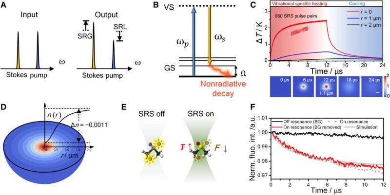
Our innovation to address this fundamental challenge is to deploy photothermal microscopy to detect the chemical bonds in a specimen. After excitation of chemical bond vibration, the energy quickly dissipates into heat, causing a rise in temperature. This photothermal effect can be measured by a probe beam passing through the focus.
Our method is fundamentally different from coherent Raman scattering microscopy, a high-speed vibrational imaging platform described in my 2015 science review. Together, we have established a new class of chemical imaging toolbox, termed vibrational photothermal microscopy, or VIP microscopy.
In the Nature Communications paper, we have developed a wide-field mid-infrared photothermal microscope to visualize the chemical content of a signal viral particle. In the Science Advances paper, we have developed a novel vibrational photothermal microscope that is based on the stimulated Raman process.
Were there any unexpected or surprising results in either paper? If so, how do these results challenge existing knowledge or theories around microscopy?
Development of SRP microscopy was unexpected. We never believed the Raman effect was strong enough for photothermal microscopy, but our thoughts shifted in August of 2021. To celebrate my 50th birthday, my students and I organized a sports-themed party. During the festivities, Yifan Zhu, the first author of the Science Advances paper, unfortunately sustained an injury, leading his doctor to recommend a two-month period of restricted mobility.
During his recovery, I asked him to run a calculation of temperature rise in the focus of an SRS (stimulated Raman scattering) microscope. Through this accident, we found a strong stimulated Raman photothermal (SRP) effect. Yifan and other students then spent two years on the development. This is how SRP microscopy was invented.
Did the papers identify any limitations or gaps in their findings? How might these limitations impact the overall implications of the research?
Certainly, nothing is perfect. In pursuing SRP microscopy, we found that each beam can have absorption, which causes a weak non-Raman background in the SRP image. We are developing a novel way to remove this background.
Do the findings of one paper complement or contradict the findings of the other? How do they relate to each other?
The methods reported in these two papers are complimentary. The WIDE-MIP method is good for detecting IR-active bonds, while the SRP method is sensitive to Raman-active bonds.
Do the papers suggest new directions for future microscopy research that could have significant long-term implications?
Yes, indeed. These two papers together indicate a novel class of chemical microscopy termed as vibrational photothermal microscopy or VIP microscopy. VIP microscopy offers a very sensitive way of probing specific chemical bonds; thus we can use them to map molecules of very low concentrations without dye labeling.
Are these imaging technologies currently available or being used by other researchers outside of your lab?
We have filed provisional patents for both technologies via BU's Technology Development office. At least two companies are interested in commercialization of the SRP technology and one of those is also interested in the WIDE-MIP technology as well.
Who are your key research collaborators?
In the WIDE-MIP paper, the virus samples are provided by John Connor, an associate professor of microbiology at BU's National Emerging Infectious Diseases Laboratories. The WIDE-MIP technology development is in collaboration with Selim Ünlü, a professor of electrical and computer engineering at BU's College of Engineering. Thus, this is a collaborative work within Boston University.
More information: Qing Xia et al, Single virus fingerprinting by widefield interferometric defocus-enhanced mid-infrared photothermal microscopy, Nature Communications (2023). DOI: 10.1038/s41467-023-42439-4
Yifan Zhu et al, Stimulated Raman photothermal microscopy toward ultrasensitive chemical imaging, Science Advances (2023). DOI: 10.1126/sciadv.adi2181
Journal information: Nature Communications , Science Advances
Provided by Boston University


