August 23, 2022 report
Sea urchin tubercules found to have a Voronoi pattern

A team of researchers affiliated with several institutions in Italy, working with a colleague from the U.S., has found that sea urchin spine joiners have bone formations that conform to a Voronoi pattern. In their paper published in Journal of the Royal Society Interface, the group describes their close-up study of the echinoids and what they learned about their spine structure.
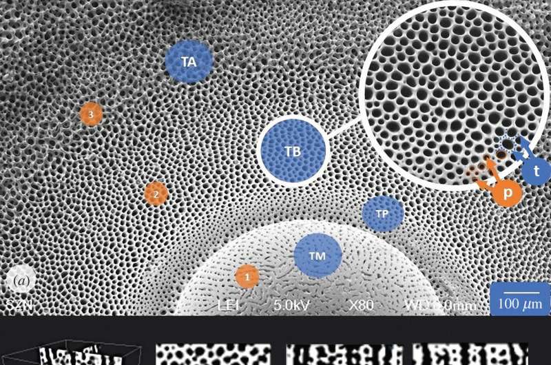
Sea urchins are spiny, usually roundish echinoderms that belong to the class Echinoidea. The spines of the sea creature are connected to a hard shell, which serves to protect the much softer organs inside. In this new effort, the researchers were studying the means by which the spines attach to the shell, via what are called skeletal tubercules. The researchers note that the tubercules must be strong to prevent predators from ripping the spines from the shell and thereby accessing the shell. To learn more about the nature of their strength, they looked at samples under a scanning tunneling microscope. The researchers discovered that the bone structure that makes up the tubercules appears to conform to a Voronoi pattern.
Voronoi patterns are created artificially using a math formula to divide a given region into polygon-shaped cells, each created around certain points called seeds. To create the pattern, which looks somewhat like a honeycomb (which also follows a Voronoi pattern, as do some dragonfly wings) cells are created following the nearest-neighbor rule, which dictates that any given spot inside of a cell is closer to its own seed than to any other seed.
To find out how closely the tubercules conform to the Voronoi pattern, the researchers took images of one section and compared it with computer-generated images that follow the pattern exactly and found an 82% match.
The researchers suggest the pattern has evolved in the tubercules to provide the sea urchin with the best possible combination of strength versus weight. They also suggest that the way the pattern has developed in the sea urchin might provide a blueprint for creating objects useful to humans—covers for sensors, for example, that are both strong and lightweight.
More information: Valentina Perricone et al, Hexagonal Voronoi pattern detected in the microstructural design of the echinoid skeleton, Journal of The Royal Society Interface (2022). DOI: 10.1098/rsif.2022.0226
Journal information: Journal of the Royal Society Interface
© 2022 Science X Network


