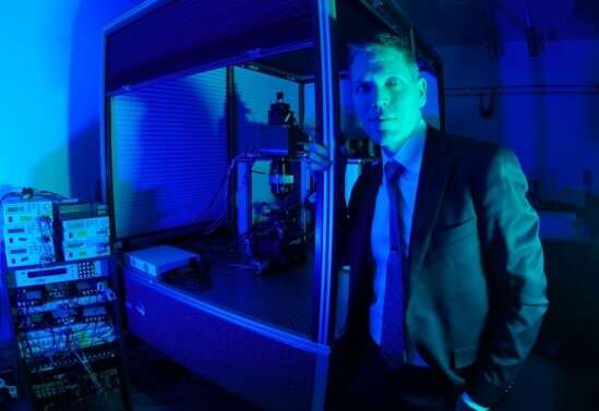
Researchers develop a two-photon microscope that provides unprecedented brain-imaging ability

Advancing our understanding of the human brain will require new insights into how neural circuitry works in mammals, including laboratory mice. These investigations require monitoring brain activity with a microscope that provides resolution high enough to see individual neurons and their neighbors.
Two-photon fluorescence microscopy has significantly enhanced researchers' ability to do just that, and the lab of Spencer LaVere Smith, an associate professor in the Department of Electrical and Computer Engineering at UC Santa Barbara, is a hotbed of research for advancing the technology. As principal investigator on the five-year, $9 million NSF-funded Next Generation Multiphoton Neuroimaging Consortium (Nemonic) hub, which was born of President Obama's BRAIN Initiative and is headquartered at UCSB, Smith is working to "push the frontiers of multi-photon microscopy for neuroscience research."
In the Nov. 17 issue of Nature Communications, Smith and his co-authors report the development of a new microscope they describe as "Dual Independent Enhanced Scan Engines for Large Field-of-view Two-Photon imaging (Diesel2p)." Their two-photon microscope provides unprecedented brain-imaging ability. The device has the largest field of view (up to 25 square millimeters) of any such instrument, allowing it to provide subcellular resolution of multiple areas of the brain.
"We're optimizing for three things: resolution to see individual neurons, a field of view to capture multiple brain regions simultaneously, and imaging speed to capture changes in neuron activity during behavior," Smith explained. "The events that we're interested in imaging last less than a second, so we don't have time to move the microscope; we have to get everything in one shot, while still making sure that the optics can focus ultrafast pulses of laser light."
The powerful lasers that drive two-photon imaging systems, each costing about $250,000, deliver ultrafast, ultra-intense pulses of light, each of which is more than a billion times brighter than sunlight, and lasts 0.0001 nanosecond. A single beam, with 80 million pulses per second, is split into two wholly independent scan engine arms, enabling the microscope to scan two regions simultaneously, with each configured to different imaging parameters.
In previous iterations of the instrument, the two lasers were yoked and configured to the same parameters, an arrangement that strongly constrains sampling. Optimal scan parameters, such as frame rate and scan region size, vary across distributed neural circuitry and experimental requirements, and the new instrument allows for different scan parameters to be used for both beams. The new device, which incorporates several custom-designed and custom-manufactured elements, including the optical relays, the scan lens, the tube lens and the objective lens, is already being broadly adopted for its ability to provide high-speed imaging of neural activity in widely scattered brain regions.
Smith is committed to ensuring open access to the instrument. Long before this new paper was published, he and his co-authors released a preprint that included the engineering details needed to replicate it. They also shared the technology with colleagues at Boston University, where researchers in Jerry Chen's lab have already made modifications to suit their own experiments.
"This is exciting," Smith said. "They didn't have to start from scratch like we did. They could build off of our work. Jerry's paper was published back-to-back with ours, and two companies, INSS and CoSys, have sold systems based on our designs. Since there is no patent, and won't be, this technology is free for all to use and modify however they see fit."
Two-photon microscopy is a specialized type of fluorescent microscopy. To perform such work in Smith's lab, researchers genetically engineer mice so that their neurons contain a fluorescent indicator of neuron activity. The indicator was made by combining a fluorescent protein from jellyfish and a calcium-binding protein that exists in nature. The approach leverages the brief, orders-of-magnitude increase in calcium that a neuron experiences when firing. When the laser is pointed at the neuron, and the neuron is firing, calcium comes in, the protein finds the calcium and, ultimately, fluoresces.
Two-photon imaging enhances fluorescence microscopy by employing the quantum behavior of photons in a way that prevents a considerable amount of out-of-focus fluorescence light from being generated. In normal optical microscopy, the light from the source used to excite the sample enters it in a way that produces a vertical cone of light that narrows down to the target focus area, and then an inverted cone below that point. Any light that is not at the narrowest point is out of focus.
The light in a two-photon microscope behaves differently, creating a single point of light (and no cones of light) that is in sharp focus, eliminating all out-of-focus light from reaching the imaging lens. "The image reveals only light from that plane we're looking at, without much background signal from above or below the plane," Smith explained. "The brain has optical properties and a texture like butter; it's full of lipids and aqueous solutions that make it hard to see through. With normal optical imaging, you can see only the very top of the brain. Two-photon imaging allows us to image deeper down and still attain sub-cellular resolution."
Another advantage of two-photon excitation light is that it uses lower-energy, longer-wavelength light (in the near-infrared range). Such light scatters less when passing through tissue, so it can be sharply focused deeper into tissue. Moreover, the lower-energy light is less damaging to the sample than shorter wavelengths, such as ultraviolet light.
Smith's lab tested the device in experiments on mice, observing their brains while they performed tasks such as watching videos or navigating virtual reality environments. Each mouse has received a glass implant in its skull, providing a literal window for the microscope into its brain.
"I'm motivated by trying to understand the computational principles in neural circuitry that let us do interesting things that we can't currently replicate in machines," he said. "We can build a machine to do a lot of things better than we can. But for other things, we can't. We train teenagers to drive cars, but self-driving cars fail in a wide array of situations where humans do not. The systems we use for deep learning are based on insights from the brain, but they are only a few insights, and not the whole story. They work pretty well, but are still fragile. By comparison, I can put a mouse in a room where it has never been, and it will run to someplace where I can't reach it. It won't run into any walls. It does this super reliably and runs on about a watt of power.
"There are interesting computational principles that we cannot yet replicate in human-made machines that exist in the brains of mice," Smith continued, "and I want to start to uncover that. It's why I wanted to build this microscope."
More information: Che-Hang Yu et al, Diesel2p mesoscope with dual independent scan engines for flexible capture of dynamics in distributed neural circuitry, Nature Communications (2021). DOI: 10.1038/s41467-021-26736-4
Journal information: Nature Communications
Provided by University of California - Santa Barbara


