How the optics of soap bubbles may help us understand the mechanics of immune cells and cancer

Scientists at the University of St Andrews have developed an advanced new microscopy technique that could revolutionise our understanding of how immune and cancer cells find their way through the body.
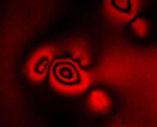
Elastic Resonator Interference Stress Microscopy (ERISM) images the extremely weak mechanical forces that living cells apply when they move, divide, and probe their environment.
As described in Nature Cell Biology today (Monday 19 June 2017), ERISM resolves the tiny forces applied by feet-like structures on the surface of human immune cells.
These feet allow immune cells to find the fastest route to a site of infection in the body. Similar structures may be responsible for the invasion of cancer cells into healthy tissue and it is planned to use ERISM in the future to learn more about the mechanisms involved in cancer spreading.
The physical effect giving soap bubbles their rainbow-like appearance is a phenomenon called thin-film interference. It is based on interaction of light reflected on either side of a soap film. The different colours that white light consists of interact with different local thicknesses of the thin film and generate the familiar rainbow patterns. Effectively the colours are an image of the film thickness at each point on the surface of the soap bubble.
A similar effect can be used to determine the forces exerted by cells. Professor Malte Gather of the School of Physics and Astronomy at St Andrews explained: "Our microscope records very high colour resolution images of the light reflected by a thin and soft probe. From these images, we then create a highly accurate map of the thickness of the probe – with a mind-blowing precision of one-billionth part of a metre.
"If cells apply forces to the probe, the probe thickness changes locally, thus providing information about the position and magnitude of the applied forces.
"Although researchers have recorded forces applied by cells before, our interference-based approach gives an unprecedented resolution and in addition provides an internal reference that makes our technique extremely robust and relatively easy to use."
This robustness means that measuring cell forces could soon become a tool in clinical diagnostics. For example, doctors may find that the ERISM method can complement existing techniques to assess the invasiveness of cancer. Work to scale up ERISM for use in the clinic is now underway.
More information: Nils M. Kronenberg et al. Long-term imaging of cellular forces with high precision by elastic resonator interference stress microscopy, Nature Cell Biology (2017). DOI: 10.1038/ncb3561
Journal information: Nature Cell Biology
Provided by University of St Andrews


