This article has been reviewed according to Science X's editorial process and policies. Editors have highlighted the following attributes while ensuring the content's credibility:
fact-checked
trusted source
proofread
Study demonstrates spontaneous self-organization of microdroplets through quasi one-dimensional confinement

Polymer systems composed of multiple components can spontaneously induce emulsion or microdroplets by mechanical mixing, as an intermediate state of macroscopic phase separation. Unfortunately, the size of generated droplets is nonuniform and their spatial-arrangement is rather random. In addition, they tend to grow larger with time (coarsening).
To prevent the change of the microdroplet size, researchers have attempted to rapidly lower the temperature, but these efforts can never improve the uniformity of the droplets. If uniformly arranged homogeneous droplets entrapping certain substrates such as DNA and medicines can be produced in a simple procedure, these droplets will serve as useful items in drug delivery and also in creating synthesis cells. This self-organization of microdroplets can provide valuable insights into the self-assembly of biological molecules.
In a study published in the journal ACS Macro Letters a research team led by Ph.D. student Mayu Shono from the Department of Chemical Engineering and Materials Science at Doshisha University, found that the homogeneous spatial pattern of microdroplets is spontaneously generated through phase separation of polymer solution along a glass capillary tube.
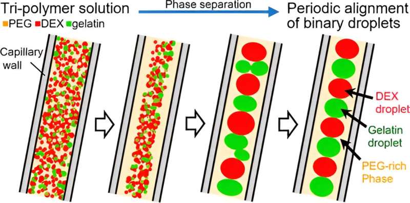
Interestingly, it was shown that the uniformly arranged pattern of the droplets are rather stable for hours. The researchers confined an aqueous tripolymer solution containing polyethylene-glycol (PEG) mixed with dextran (DEX) and gelatin in a glass capillary tube coated with PEG. They observed that over time, the three phases separated, and the DEX and gelatin droplets aligned themselves in a periodic pattern in the PEG phase.
The spontaneous self-assembly arrangement occurred without any exchange of materials or energy in the system, setting it apart from other systems. "We have been performing our study to make clear the underlying mechanism of self-organization in living matter. As a result of this study, we have discovered a novel phenomenon for the generation of self-organized characteristic pattern," says Ms. Shono.
In their experiments, the researchers prepared three aqueous tripolymer solutions, combining PEG, DEX, and gelatin with distilled water in a 5:4:6 weight ratio.
To distinguish the molecules, they labeled the DEX and gelatin with fluorescent markers. These markers emit light of specific colors when exposed to light of certain wavelengths, allowing them to identify the different components in the sample. The solution was then drawn into PEG-coated capillary tubes with diameters of 140 μm and 280 μm.
Due to preferential attachment to the capillary's surface, PEG separated from the solution immediately. The DEX and gelatin phases, which were repelled from the inner wall, then formed droplets that increased in size.
In 40 seconds, the DEX droplets formed a linear arrangement in the center of the capillary, and 120 seconds later, the gelatin droplets did the same. This led to a self-organized, periodic alignment of DEX- and gelatin-rich microdroplets surrounded by a PEG-rich phase, which maintained itself for eight hours after formation.
The essentials of the observed pattern are reproduced through numerical simulation by modifying the theoretical model with the Cahn-Hilliard equation, which describes the time-dependent change of the spatial pattern of phase separation in a mixture of three different polymers.
Achieving stable micropatterns through phase separations is challenging, because, in general, the microdroplets generated through phase transition are nonuniform and they tend to collapse or disappear with time. However, by confining them in a capillary with suitable chemical modification of its inner surface, the researchers were able to preserve the patterns for long periods.
"The novel methodology to obtain uniform droplets reported here is superior to current microfluidics in several aspects," says Ms. Shono.
In the future, such micropatterns can be studied to provide insights into the mechanisms involved in the self-assembly of biological molecules. Furthermore, it can aid in the development of targeted drug delivery and the production of desired macromolecules, such as proteins and nucleotides using protocells.
Recently, Ms. Shono together with collaborators, published an article in the journal Small indicating the successful selective entrapment of genome-sized DNA into the homogenous, arranged droplets.
Ms. Shono concludes, "This scenario for pattern formation coupled with phase separation under confinement may provide a novel viewpoint to uncover the hidden factors for the origin of life and also to reveal the underlying mechanism on the stability of the structure and function of membrane-less organelles in living cells."
More information: Mayu Shono et al, Periodic Alignment of Binary Droplets via a Microphase Separation of a Tripolymer Solution under Tubular Confinement, ACS Macro Letters (2024). DOI: 10.1021/acsmacrolett.3c00689
Provided by Doshisha University


