This article has been reviewed according to Science X's editorial process and policies. Editors have highlighted the following attributes while ensuring the content's credibility:
fact-checked
peer-reviewed publication
proofread
Researchers identify molecular basis for morphological diversity of amniote skull

A research group led by Associate Professor Masayoshi Tokita explored the molecular basis generating the diversity of amniote skull morphology, using embryos of several amniote species as materials.
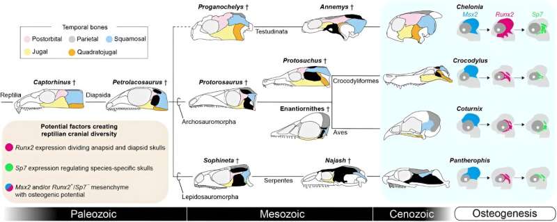
Their study suggests that the diversity of amniote skull morphology may be brought about by spatiotemporal differences in the expression of three osteogenic genes, Msx2, Runx2, and Sp7.
The finding provides a basis for understanding how skull morphology has diversified in amniotes, the first vertebrate lineage to fully move to land, including humans. Furthermore, the findings may provide hints for the development of treatments for congenital diseases that cause abnormalities in human skull morphology. The findings of this research are reported in Science Advances.
Amniote skulls have an opening(s) surrounded by bones behind the orbit (temporal region), called the temporal fenestration(s). Amniote skulls are categorized into three conditions: anapsid, synapsid, and diapsid, depending on the number of temporal arches that border the ventral side of the temporal fenestrations.
In this study, the expression patterns of the osteogenic genes Msx2, Runx2, and Sp7 were examined during embryonic skull formation of six reptilian species (three turtle species, two snake species, and one crocodile species), two avian species, and two mammalian species. In the temporal region of reptilian and avian embryos, Msx2 was widely expressed.
The expression domain of Runx2 was widespread in the temporal region of turtle embryos, whereas in non-turtle reptiles and birds, it was restricted to the region excluding the domain where the temporal fenestrations later appeared
In the temporal region of reptilian and avian embryos, Sp7 was regionally expressed in the area where the bones are later formed. Its expression pattern varied among species. In the temporal region of mammalian embryos, Msx2, Runx2, and Sp7 were regionally expressed in the domain where the bones were formed.
This study suggests that the diversity of amniote skull morphology may be brought about by spatiotemporal differences in the expression patterns of Msx2, Runx2, and Sp7.
More information: Hiromu Sato et al, Turtle skull development unveils a molecular basis for amniote cranial diversity, Science Advances (2023). DOI: 10.1126/sciadv.adi6765
Journal information: Science Advances
Provided by Toho University


