This article has been reviewed according to Science X's editorial process and policies. Editors have highlighted the following attributes while ensuring the content's credibility:
fact-checked
peer-reviewed publication
trusted source
proofread
3D organoids unlock promising insights into lung cancer in dogs

Veterinary researchers have used organoids—three-dimensional organ-like structures grown from stem cells and tissue samples—to investigate the biological processes of lung cancer in dogs, a disease that is much rarer in our canine friends than it is in humans, but often far more deadly
Organoids are three-dimensional organ-like structures grown in the lab that offer far greater fidelity to complex biological processes than traditional cell cultures grown on flat 2D surfaces. These cutting-edge models have only recently been developed to aid research into human biology and disease. But researchers have now for the first time applied the research technique to lung cancer in dogs.
A study describing the researchers' techniques and findings was published in the journal Biomedicine & Pharmacotherapy.
In the realm of cancer research, lung cancer has always loomed as a formidable adversary in the human world, but our beloved canine companions can also suffer from their own version of this disease, known as canine primary lung cancer.
For dogs diagnosed with lung cancer, the outlook can be grim. Many cases are only discovered when the disease has already progressed significantly, leading to limited treatment options. Surgical removal of lung tissue, known as lung lobectomy, is a common approach, but it may not be curative for advanced cases.
In fact, the median survival time for dogs undergoing lung lobectomy hovers between 160 and 450 days, even with chemotherapy. For dogs with extensive cancer growth, the prognosis is even bleaker, with survival times ranging from a mere 60 to 180 days.
Unlike human medicine, where molecular targeted therapies have become a game-changer, such treatments are scarce in veterinary practice, leaving our furry friends with limited options.
Enter the world of 3D organoid cultures, a cutting-edge technology that mimics the dynamics of living tissue better than traditional cell cultures. 3D organoids are miniature, three-dimensional structures that are cultured from stem cells or tissue samples in a lab. The stem cells or tissue samples can self-organize into complex, organ-like structures—hence "organoid."
The organoid's cells and structures can interact with each other just like in a real organ in a living organism in three dimensions, instead of traditional cell cultures grown on flat, 2D surfaces. This allows scientists to study much more complex biological processes.
The veterinary medicine researchers thought that they could apply this new technique to the thorny problem of canine lung cancer in the hope that they could identify novel therapeutic avenues.
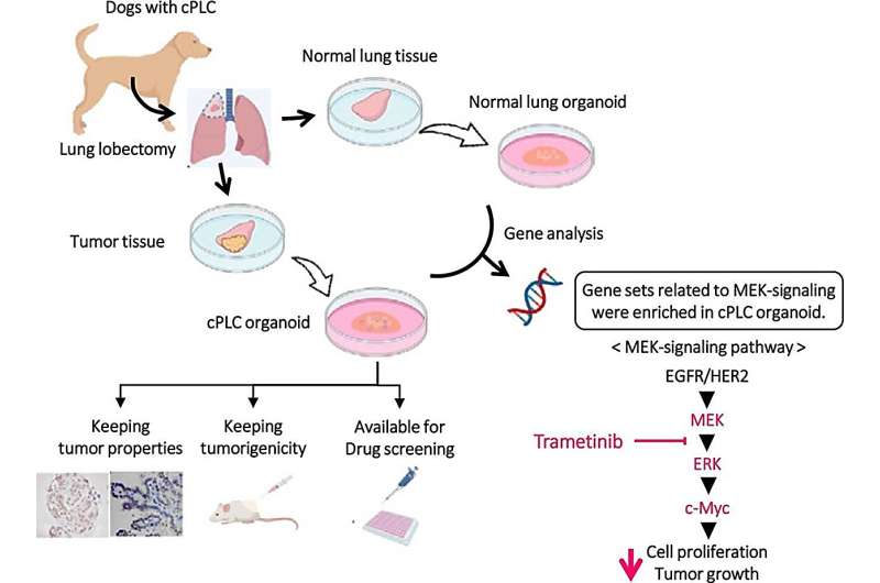
They took samples of tumors from dogs with lung cancer as well as from the healthy parts of their lungs to create canine primary lung cancer organoids (cPLCO) and canine normal lung organoids (cNLO).
"The hope was that these organoids would replicate the tissue architecture of dog lung cancer better than any previous attempts at cell cultures," said Tatsuya Usui corresponding author of the study and Associate Professor at the Division of Animal Life Science with Tokyo University of Agriculture and Technology.
The researchers found that the cPLCO faithfully mirrored the characteristics of their original tumor tissues, both in histological morphology (in essence, the shape and structure of the tissues) and molecular profiles, including the expression of specific tumor markers. Tumor markers are any molecules, typically proteins, produced by the cancer cells, or by other cells in the body, in response to the presence of a cancer that give any information about the cancer.
These markers, produced in higher amounts than in normal healthy patients, are what clinicians and researchers use to identify how aggressive the cancer is and whether it is responding to treatment.
They found that different strains of canine lung cancers exhibited varying sensitivity to anti-cancer drugs, opening the door to the potential for personalized treatment approaches.
The scientists also identified a correlation between the viability of organoid cells and the expression levels of specific target molecules (basically how often a gene gets "switched on"), such as Human Epidermal Growth Factor Receptor 2 (HER2) and Epidermal Growth Factor Receptor (EGFR). These are both proteins that play crucial roles in cell signaling, growth and regulation. This observation could pave the way for therapies that target these molecules in the future.
Sequencing of RNA revealed that the cancerous organoid exhibited significant increase in the "switching on" of 11 genes associated with tumor proliferation in other cancers.
Perhaps most tantalizingly, the cancerous organoid displayed enrichment in the Mitogen-Activated Protein Kinase signaling pathway, also known more simply as the MEK-signaling pathway, a promising avenue for intervention, as its molecular processes enable transmission of information from receptors on the surface of a cell to the DNA in its nucleus.
This pathway, involving a cascade of proteins acting on other proteins, in turn acting on other proteins, and so on, is key to cell proliferation, differentiation, survival, and responding to information signals from outside the cell. Often, if any one of the steps on the process gets stuck in an on or off position, it can give rise to tumor growth.
The researchers tested a MEK inhibitor called trametinib, which significantly decreased the viability of the cancerous organoid and inhibited tumor growth when cancer cells were grafted onto healthy cell models.
The creation of dog lung cancer organoids provides a powerful tool to explore the disease in unprecedented detail, potentially leading to more effective treatments for our four-legged friends—but may even extend its influence into the realm of human lung cancer, offering new diagnostic markers and therapeutic targets.
More information: Yomogi Shiota (Sato) et al, Derivation of a new model of lung adenocarcinoma using canine lung cancer organoids for translational research in pulmonary medicine, Biomedicine & Pharmacotherapy (2023). DOI: 10.1016/j.biopha.2023.115079
Journal information: Biomedicine & Pharmacotherapy
Provided by Tokyo University of Agriculture and Technology


