June 26, 2023 report
This article has been reviewed according to Science X's editorial process and policies. Editors have highlighted the following attributes while ensuring the content's credibility:
fact-checked
peer-reviewed publication
trusted source
proofread
Cochlea cell atlas built from single-cell sequencing discovers new cell types, uncovers hidden molecular features

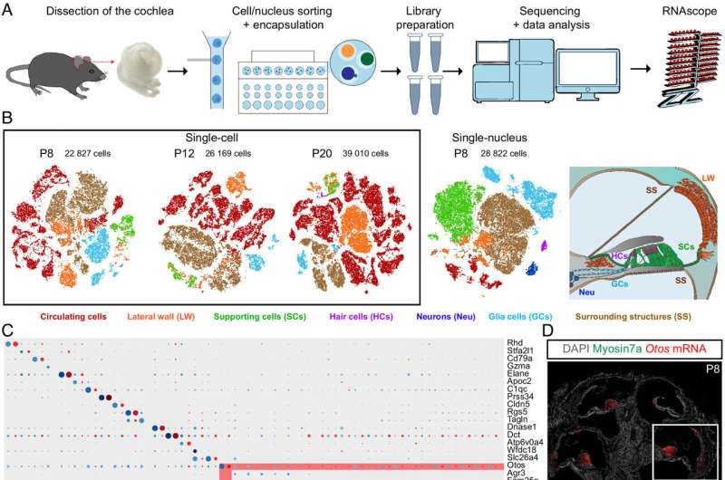
Researchers at the Pasteur Institute in France have conducted an in-depth genomic study of mouse cochlea to create a comprehensive transcriptomic atlas of the auditory organ at a molecular level.
In their paper, "Single-cell transcriptomic profiling of the mouse cochlea: An atlas for targeted therapies," published in the Proceedings of the National Academy of Sciences, the team details the creation of a genomic database for understanding the gene regulatory networks involved in cochlear cell differentiation and maturation, which are essential for creating targeted treatments.
The study analyzed more than 120,000 cells and characterized their gene expression profiles. The resulting cell atlas provides information about nearly all cochlear cell types and identifies cell type–specific markers. Three previously unknown cell types were discovered, two contributing to the modiolus and one to cells lining the scala vestibuli.
The two previously unknown modiolus-related cells are transcriptomically distinct from other modiolus cell types, and the researchers think they may be related to the spongious structure of the modiolar bone. They also state that it is possible that the two cells are actually one cell type at different stages of maturity, as known modiolar bone cells do have distinct maturity roles in bone formation.
The third cell type discovery was found bordering the scala vestibuli, covering the epithelial cells of Reissner's membrane, and are being called scala vestibuli border (SVB) cells. SVB cells expressed several causal genes for various deafness forms, including Eps8l2, Gjb2, Gjb6, Homer2, Coch, Clic5, Dcdc2a, Pou3f4, Col4a6, and Six1. This cell tissue has never been identified before in any histological studies.
The study also sheds light on the molecular basis of the tonotopic gradient of the basilar membrane's biophysical characteristics, the main mechanical element of the inner ear crucial for sound frequency analysis.
The basilar membrane varies in width, thickness, and stiffness progressively along the length of the cochlea and operates as a frequency analyzer, generating a tonotopic map with high-frequency sounds detected at the base and low-frequency sounds at the apex of the cochlea.
The stiffness gradient of the basilar membrane is thought to depend on the Emilin-2 protein, which contributes to extracellular functions and tissue elasticity and is synthesized by tympanic border cells. Emilin2 mRNA was revealed to have a gradient of expression along the membrane, with the intensity decreasing from base to apex, confirming the previous expectations.
Emilin2 was not alone, and several transcription factors also displayed a tonotopic intensity gradient similar to that of Emilin2 (Gata6, Cux2, Nr1h3, Atoh8, and Atf3), while others followed gradients in the opposite direction (Sp5, Foxf2, Dach1, Pbx3, Tbx1, Creb5, Osr1, Zic2, and Zic5).
The expression of deafness genes in several cochlear cell types was revealed. Researchers analyzed cell expression patterns of 120 detected genes involved in isolated forms of deafness and 75 detected essential genes for cochlear development and function.
Deafness genes showed low expression levels in some cochlear cell types, increasing only in a given cochlear cell type at a particular stage. This manifestation of deafness gene expression suggests a need to design therapeutics specifically targeting certain combinations of cell types at appropriate time points.
This new single-cell sequenced atlas will be handy for finding overlooked cochlear cells affected by particular deficits and may reveal causal sources for deafness of unknown origin. Future research can also use the reference in deciphering cell signaling pathways and gene regulatory networks to develop safe and effective gene therapies.
More information: Philippe Jean et al, Single-cell transcriptomic profiling of the mouse cochlea: An atlas for targeted therapies, Proceedings of the National Academy of Sciences (2023). DOI: 10.1073/pnas.2221744120
Journal information: Proceedings of the National Academy of Sciences
© 2023 Science X Network


