How growth factor Midkine suppresses AMPK during cancer progression

A research group led by Prof. Piao Hailong from the Dalian Institute of Chemical Physics (DICP) of the Chinese Academy of Sciences (CAS) uncovered a new function of growth factor Midkine to suppress the Liver Kinase B1 / AMP-Activated Protein Kinase (LKB1-AMPK) axis in a noncanonical intracellular manner.
They found that by disrupting the LKB1-STRAD-Mo25 (STRAD: STE20-Related Kinase Adaptor) complex, Midkine decreased LKB1 activity and attenuated AMPK phosphorylation to promote cancer cell proliferation and tumor progression.
This study was published in Cell Death and Disease on April 29.
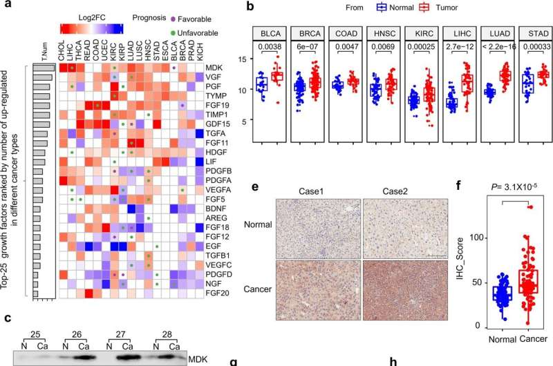
Midkine belongs to the pleiotrophin family growth factor and participates in diverse physiological processes. The expression of Midkine is upregulated in different types of human malignancy.
Like other growth factors, Midkine is secreted to extracellular space after signal peptide cleavage. Usually, the secreted Midkine is considered as a ligand, which binds to different transmembrane receptors to induce intracellular signaling.
Although a few studies have reported that the secreted Midkine can be transported back into cells through endocytosis, it is still unclear whether intracellular Midkine performs biological functions.
In this study, the researchers found that the relocalization of Midkine was highly efficient, and the internalized Midkine was mainly detected in the cytoplasm, indicating some unreported intracellular function of Midkine.
The researchers focused on AMPK signaling and found that Midkine suppressed AMPK phosphorylation at the Thr172 site of the α subunit. This resulted in the inactivation of AMPK in an intracellular dependent manner, and when Midkine was detained in extracellular space by heparin, the repression of Midkine to AMPK was relieved.
Furthermore, they found that Midkine suppressed AMPK activation through the upstream kinase LKB1. Mass spectrometry (MS) analysis combined with immunoprecipitation test proved that Midkine was associated with LKB1 and STRAD to disrupt the LKB1-STRAD-Mo25 complex, which was indispensable for LKB1 activation.
By engineering the expression of Midkine and LKB1, the researchers verified that Midkine promoted cancer cell proliferation through the LKB1-AMPK axis. The analysis of clinical data also confirmed that the expression of Midkine was negatively related to AMPK activity and patient prognosis.
More information: Tian Xia et al, Midkine noncanonically suppresses AMPK activation through disrupting the LKB1-STRAD-Mo25 complex, Cell Death & Disease (2022). DOI: 10.1038/s41419-022-04801-0
Journal information: Cell Death and Disease
Provided by Chinese Academy of Sciences


