January 24, 2022 feature
SARS-CoV-2 spike protein activates human endogenous retroviruses in blood cells

Transposable elements, or jumping genes, are now known to be responsible for many human diseases. Keeping them repressed by methylation, RNA binding, or the attentions of the innate immune system is a full-time jump for cells.
Last week, we reviewed the activation of one particular kind of transposable element, the Line-1 retrotransposons, in an ever-expanding host of neurodegenerative conditions. Retrotransposons derive from human endogenous retrovirus (HERVs) but typically have lost their signature long terminal repeat sequences at the beginning and ends of their genes.
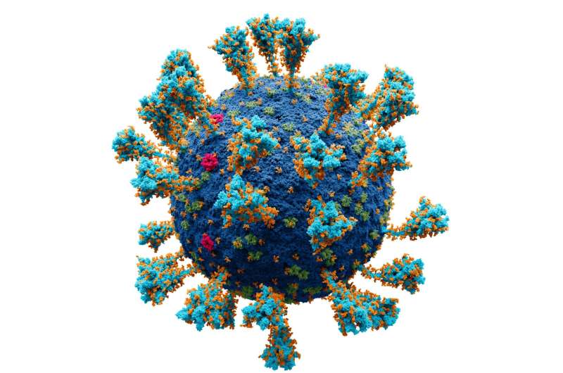
On Tuesday, a real zinger was dropped onto the medRxiv preprint server that could potentially explain many of the commonly observed pathogenic features of SARS-CoV-2. The authors provide solid evidence that the SARS-CoV-2 spike protein activates the envelope (ENV) protein encoded by HERV-W in blood cells, which is in turn directly responsible for many pathological features of the disease. HERV-W is named for the fact that many retroviruses in the group use a tryptophan tRNA in the primer binding site. Apparently, the shape of the letter W somehow reminded the naming committee of the shape of the ring structure of atoms in the side chain of tryptophan.
Researchers had previously observed a correlation in the expression of HERV-W ENV protein in T lymphocytes with severe respiratory distress in SARS-CoV-2 patients. However, the exact mechanisms involved were not clear. Now, the real culprit in HERV-W activation has been discovered. Researchers added a recombinant trimeric spike protein without stabilizing mutations to cultured peripheral blood mononuclear cells (PBMCs) from SARS-CoV-2 patients. They found immediate and significant upregulation of the RNAs for the ENV protein from both HERV-W and HERV-K. Curiously, only the RNAs for HERV-W resulted in subsequent ENV protein expression.
Native spike proteins tend to prematurely refold into a post-fusion conformation, which compromises immunogenic properties and prefusion trimer yields. mRNA vaccines therefore have slight modifications that simultaneously make the mRNA less immunogenic, and the spike protein it encodes more immunogenic. One way this has been done is to stabilize specific conformers through the addition of two strategic prolines to the code. However, more research is needed to fully characterize the fusogenic potential of stabilized spike proteins. Some vaccine manufacturers have eliminated the furin cleavage site from their mRNA construct in order to reduce potential residual fusion of a 2-PP stabilized construct. A few of these observations were initially pointed out to me by an anonymous researcher on social media operating with the moniker "Underground courtlady."
A key finding in these studies is that not all COVID patients had significant HERV-W ENV activation; only 20 or 30 percent of them did. This finding likely reflects an underlying genetic susceptibility among the infected that absolutely needs to be defined and taken into account, particularly if HERV-W is going to be used as a general marker for disease severity, or as a therapeutic target for a humanized monoclonal antibody therapy, as is now envisioned. For example, activation of a soluble hexameric form of HERV-W was found in multiple sclerosis, and is earmarked as potentially druggable option.
But which HERV-W, exactly? Over 1 percent of our genomes are HERV-W remnants, more than all our protein-coding regions put together. In fact, there are at least 13 HERV-W loci with full-length ENV genes in the human genome. One of these, which hails from chromosome region 7q21.2, has an uninterrupted open reading frame for a complete HERV-W ENV protein. This protein, Syncytin-1, figures famously and essentially in normal placental development. To complicate things, MS now seems to have many eclectic potential origins. Researchers revealed this week, to considerable acclaim, that infection with Epstein-Barr virus is an important upstream, or downstream, or perhaps altogether independent trigger for MS.
HERV-W is not the only retroviral game in town. Researchers recently discovered that a retrovirus-like protein known as PEG10 directly binds to and secretes its own mRNA in extracellular virus-like capsids. This behavior is eerily similar to that of the ARC1 retroviral protein now understood to be critical in the formation of memory at synaptic sites. Incredibly, researchers are already well on their way to pseudotyping these virus-like particles with fusogens to create an endogenous vector for delivering functional mRNA cargos as a gene therapy. Clearly, some caution in these affairs is warranted (warning: opinion).
In heart tissue samples from COVID-19 patients, HERV-W ENV was mainly found in endothelial cells from numerous small blood vessels and in the pericardial fatty tissue. The endothelial nature of HERV-W ENV positive cells was confirmed in this case with CD31 staining. Ominously, significant HERV-W ENV in patients was found in blood clots, nasal mucosa and also in the central nervous system, particularly in microglial cells, even when SARS-CoV-2 could not be detected in those tissues. The authors note that SARS-CoV-2 induced HERV-W ENV expression in human lymphoid cells, cells that neither express the canonical ACE2 receptor, nor the TMPRSS2 protease. This suggests other routes for the virus into these cells. One recent clue to other candidate mechanisms might come from alternative receptors like ASGR1, which is highly expressed in liver cells.
It is now of the utmost importance to find out how SARS-CoV-2 activates HERVs. In light of the known penchant for transposable elements to both be activated by, and further integrate into sites of active DNA repair, it may be worth revisiting earlier studies that purported to show that reverse transcribed SARS-CoV-2 RNA could integrate into the genome of cultured human cells and subsequently express in patient-derived tissues. These authors found target site duplications flanking the viral sequences and consensus LINE1 endonuclease recognition sequences at the integration sites—features consistent with a LINE1 retrotransposon-mediated, target-primed reverse transcription and retroposition mechanism.
More information: Benjamin Charvet et al, SARS-CoV-2 induces human endogenous retrovirus type W envelope protein expression in blood lymphocytes and in tissues of COVID-19 patients, medRxiv (2022). DOI: 10.1101/2022.01.18.21266111
© 2022 Science X Network


