Insight into the evolution of bones

A joint team of paleontologists has now for the first time analyzed bone structures in 400 million-year-old fossils of marine life at unprecedentedly high resolution and in 3D. To be able to view these structures, tomography experts at the Helmholtz-Zentrum Berlin (HZB) examined the samples under the focused ion beam of a scanning electron microscope to calculate 3D images from the data, achieving resolutions in the nanometre range using technology that was initially developed to study battery corrosion.
Whether fish, fowl or mammal, all vertebrates have an internal skeleton of bones. In almost all vertebrates (with the exception of certain bony fish), the bone consists of a complex composite of minerals, proteins, and living bone cells (osteocytes) entrapped in the bone matrix. The bone cells are interconnected by tiny channels so that they can exchange substances and electrochemical signals, allowing the bone to grow and regenerate. Still, this complex architecture of live and inorganic material must have emerged at some point in the course of evolution. A team at the Museum für Naturkunde Berlin headed by Dr. Florian Witzmann is investigating how and when this happened. Now they have discovered a possible milestone in this development.
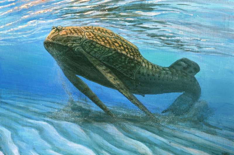
They have examined fossilized samples of bony armor from two early fish species that lived around 400 million years ago. One sample came from Tremataspis mammillata, a jawless fish that lived in the late Silurian period about 423 million years ago and belongs to the extinct group called Osteostraci. The second, much younger sample was a piece of bone from the fish Bothriolepis trautscholdi that lived in the late Devonian period about 380 million years ago and belongs to the extinct Placodermi, the earliest group of jawed fishes. "It was already known that these early vertebrates had bone cells, but we knew little about how the cells were connected to each other, as well as anything about the detailed structure of the lacuna, or cavities, in which the bone cells were located in the living animal. In order to be able to make more precise statements about bone metabolism, we had to have far more detailed images of these structures than were previously available," says Witzmann.
To achieve this, HZB expert Dr. Ingo Manke suggested a method that is available at the HZB campus in Wannsee in the Electron Microscopy Laboratory: focused ion-beam scanning electron microscopy (FIB-SEM) tomography on the ZEISS Crossbeam 340. In this device, a focused gallium-ion beam continuously ablates material from the sample surface, gradually digging its way deeper into the sample. At the same time, an electron beam scans the freshly revealed part of the sample and provides data for creating 3D images at a resolution that is more than a hundred times finer than computer tomography.

Manke's team had already used this method to study electrode materials for batteries, which have a network of fine paths for transporting ions. HZB physicist Markus Osenberg had previously employed a sophisticated evaluation procedure developed at HZB's 3D Analytics Lab to calculate the image from the measurement data. This is a specially trained neural network, a method borrowed from machine learning, because images of these kinds of samples cannot be calculated using standard methods. "Due to the countless paths through the bone, the sample surface is as full of holes as Swiss cheese," explains Osenberg, who is doing his doctorate in Manke's team. However, after some practice, the well-trained neural network recognizes where the plane of the ablation runs and where the holes are, and reconstructs an accurate image of the ablated surface. "In fact, the structures in the bone samples are relatively similar to the structures in the electrode materials of batteries. But the fact that the neural network, which learned on battery materials, can now also image the fossil bone samples so well surprised us," says Osenberg.
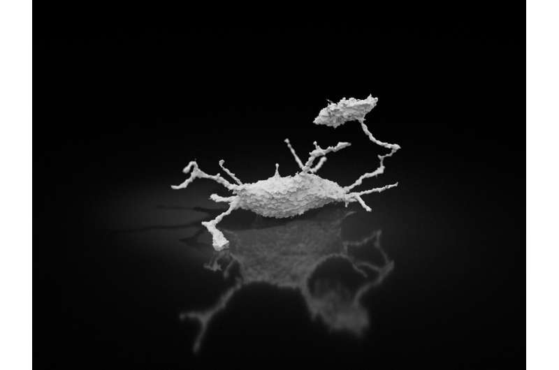
Even in the older sample of the jawless armored fish, the 3D images display a complex network with cavities (lacunae) for the bone cells and tiny channels through the bone interconnecting these cavities. "The channels are a thousand times narrower than a human hair and yet, amazingly, they have been almost completely preserved over these 400 million years," says Manke.
Elaborate analysis of the high-resolution 3D images shows in detail how the network was constructed of cavities (lacuna) and the channels between them. "This proves that our early, still-jawless ancestors already possessed bones characterized by internal structure similar to ours and probably by many similar physiological capabilities as well," Witzmann explains. "The most important palaeobiological finding is that we can also detect actual traces of metabolism in these earliest bone samples," says Yara Haridy, who is doing her Ph.D. at the Museum für Naturkunde Berlin. Through local osteolysis, i.e. dissolution of the bone matrix that surrounded the bone cells, the organism was probably able to cover its need for phosphorus in times of scarcity. This gave it an advantage over its more primitive contemporaries, who had cell-free bone, i.e. whose bones contained no osteocytes.
"This advantage apparently led to the widespread establishment of bones with bone cells in vertebrates, as we know it in humans as well. It is an important step towards understanding how our own bone metabolism came about," Haridy explains. "Even in early fossil bone, bone cells could dissolve and restore bone minerals, this means that bones themselves act as batteries by storing minerals and releasing them later! This ability provided an undoubtable advantage to jawless fish with bone cells over vertebrates without. This advantage was possibly so profound as to alter vertebrate evolution, as later jawed vertebrates retained bone cells."
The study is published in Science Advances.
More information: "Bone metabolism and evolutionary origin of osteocytes; novel application of FIB-SEM tomography" Science Advances (2021). DOI: 10.1126/sciadv.abb9113
Journal information: Science Advances
Provided by Helmholtz Association of German Research Centres


