The tug-of-war at the heart of cellular symmetry

Symmetry and asymmetry are fundamental properties of nature. Seen from above, butterflies have left-right symmetry, while male fiddler crabs show dramatic asymmetry. This is also the case for the fundamental units of life: cells. They control the symmetry of their internal structures to regulate all biological functions.
Publishing in Nature Communications, a team led by Kyoto University's Hakubi Center for Advanced Research has announced the development of an artificial cell that brings to light the dynamics that govern each cell's internal symmetry.
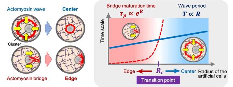
The team found that the actomyosin network—a complex consisting primarily of the protein actin that creates the filamentary meshwork, and the force-generating molecular motor myosin—self-organizes into two distinct structures that push-and-pull intracellular components as if in a tug-of-war.
Previous studies have shown that the structural mass of a cell—the actin cytoskeleton—is involved in symmetrical positioning. It has been hypothesized that this network steers and positions intracellular components. However, the mechanisms of how the proteins find the 'center' of the cell, or how they induce symmetry breaking, have remained elusive.
"Living cells are traditionally used to study these processes," explains Makito Miyazaki who led the study. "But a cell is so complex that it can obfuscate the underlying regulatory system."
To overcome this difficulty, the team settled on a bottom-up approach, developing a simplified, artificial cell by confining the actin cytoskeleton in a tiny droplet of liquid. This enabled them to control the sizes and concentrations of any proteins of interest.
By then changing the cell size, the team discovered two coexisting actomyosin networks with opposing functions: a ring-like centripetal actomyosin that pushes toward the center, and radially-formed bulk actomyosin bridges that pull to the edges.
Molecular perturbation experiments and theoretical modeling further revealed that the balance between these two networks is what determines positioning symmetry.
"How cells organize their internal structures is an important question, which we must answer in order to understand how our bodies are constructed from single fertilized cells," concludes Miyazaki.
By simplifying the cell system, the team believes that it has developed a universally-applicable model that could lead to further revelations regarding life's most fundamental functions.
More information: Ryota Sakamoto et al, Tug-of-war between actomyosin-driven antagonistic forces determines the positioning symmetry in cell-sized confinement, Nature Communications (2020). DOI: 10.1038/s41467-020-16677-9
Journal information: Nature Communications
Provided by Kyoto University


