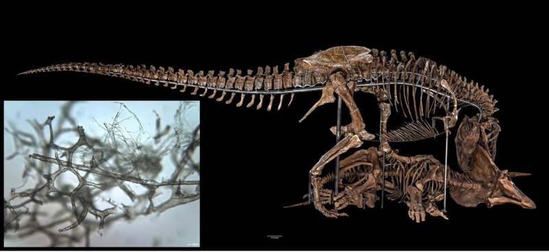
How dinosaur blood vessels are preserved through the ages

A team of scientists led by Elizabeth Boatman at the University of Wisconsin Stout used infrared and X-ray imaging and spectromicroscopy performed at Berkeley Lab's Advanced Light Source (ALS) to demonstrate how soft tissue structures may be preserved in dinosaur bones—countering the long-standing scientific dogma that protein-based body parts cannot survive more than 1 million years.
In their paper, now published in Scientific Reports, the team analyzed a sample from a 66-million-year-old Tyrannosaurus rex tibia to provide evidence that vertebrate blood vessels—collagen and elastin structures that don't fossilize like mineral-based bone—may persist across geologic time through two natural, protein-fusing "cross-linking" processes called Fenton chemistry and glycation.
First, the scientists used imaging, diffraction, spectroscopy, and immunohistochemistry to establish that structures present in the sample are indeed the animal's original collagen-based tissue. Then, Berkeley Lab co-authors Hoi-Ying Holman and Sirine Fakra respectively performed synchrotron radiation-based Fourier-transform infrared spectromicroscopy (SR-FTIR) to examine how the cross-linked collagen molecules were arranged, and X-ray fluorescence (XRF) mapping to analyze the distribution and types of metal present in T. rex vessels.
"SR-FTIR takes images and spectra of the same sample, and so you can reveal the distribution of protein-folding patterns, which helps to identify the possible cross-linking mechanisms," said Holman, director of the Berkeley Synchrotron Infrared Structural Biology (BSISB) Imaging Program. Fenton chemistry and glycation are both non-enzymatic reactions—meaning they can occur in deceased organisms—that are driven by the iron present in the body.
"The XRF microprobe revealed the presence of finely crystalline goethite, a very stable iron oxyhydroxide mineral, on the vessels that likely contributed to the preservation of organic molecules," said Fakra, an ALS research scientist.
The authors believe that the cross-linking reactions they found evidence of, combined with the protection offered from being surrounded by dense mineralized bone, can explain how original soft tissues persist.
More information: Elizabeth M. Boatman et al. Mechanisms of soft tissue and protein preservation in Tyrannosaurus rex, Scientific Reports (2019). DOI: 10.1038/s41598-019-51680-1
Journal information: Scientific Reports
Provided by Lawrence Berkeley National Laboratory


