Innovative imaging technology leads to automated pathological diagnosis

FUJIFILM Corporation has developed surface-enhanced Raman spectroscopy (SERS) imaging technology capable of analyzing large areas of unlabeled/unstained tissue metabolites with high precision using SERS that enhances the Raman scattering light when the target substance is irradiated with light, detecting substances with a high sensitivity.
In joint research conducted by Fujifilm's Frontier Core-Technology Laboratories, Research and Development Management Headquarters and Keio University (Visiting Professor Makoto Suematsu, President of the Japan Agency for Medical Research and Development as a main role, Yasuaki Kabe, full-time lecturer of Department of Medical Chemistry, School of Medicine, and others), this technology succeeded in the world's first automated visualization of the distribution of cancer tissues in a section of mouse tissues from metabolite information from SERS imaging. These are innovative results that will lead to the automated pathological diagnosis of cancers, such as resistance to anti-cancer agents and determination of the degree of malignancy, in addition to enable more accurate discrimination of the progression of cancers. The results of this research will be published in the online highlight edition of the scientific journal Nature Communications on April 19, 2018.
For analyzing cancerous tissues and lesions, methods such as labeling with antibodies and imaging of cell morphology through staining are being commonly used in terms of methods. Analyzing the slight morphological abnormalities of a cell with high precision, however, requires considerable skill. In this context, imaging using SERS (SERS imaging) is drawing attention for its ability to analyze tissues by identifying substances such as metabolites that are unique to the lesion rather than conducting a morphological analysis using labeling and staining.
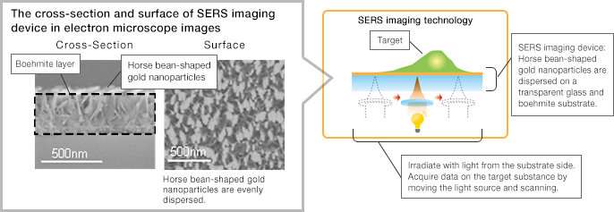
SERS imaging is an analytical method that uses a phenomenon called Raman scattering, in which light with wavelength unique to the molecular structure is scattered when the target substance on a substrate is irradiated with light. In addition, by placing metal microparticles such as nano-sized gold on the substrate, the Raman scattering from the target substance can be enhanced, enabling the detection of substances with high sensitivity. However, it was difficult to place metal microparticles evenly over a large area of the substrate and to extract only the necessary information from the enhanced Raman scattering to create an image.
At its Frontier Core-Technology Laboratories, Fujifilm utilized its grain formation technology and nanophotonics technology cultivated through the photographic film business to develop SERS imaging technology capable of analyzing large areas of unlabeled/unstained tissue metabolites with high precision.

Characteristics of SERS imaging technology developed by Fujifilm
Capable of detecting the target substance in a large area with sensitivity significantly higher than the conventional Raman imaging through a design that utilizes the grain formation technology and nanophotonics technology, dispersing horse bean-shaped gold nanoparticles evenly over the substrate.
Capable of imaging the target substance such as the lesion precisely by extracting only the necessary information with a proprietary imaging algorithm.
Light can be irradiated from the substrate side by using transparent glass and boehmite as the substrate. As light can be irradiated and the Raman scattering can be detected without being obstructed by the target substance, highly precise analysis is possible.
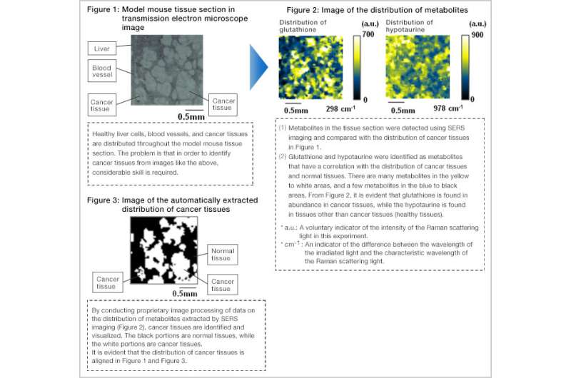
Further, in the joint research conducted with the Keio University School of Medicine, Department of Chemistry, the distribution of cancer tissues in the model mouse tissue sections and the Raman scattering of the respective metabolites detected with SERS imaging technology were compared, and metabolites with the distribution unique to cancer were identified. In addition, the study succeeded in the world's first automated visualization of the distribution of unlabeled/unstained cancer tissues through the SERS imaging with proprietary image processing. By advancing this technology, there is the possibility that it may lead to more accurate discrimination of the progression of cancers from metabolite information, rather than diagnoses based on slight morphological abnormalities. Moreover, it is expected to lead to the realization of qualitative diagnoses of cancer such as determining whether there is resistance to anti-cancer agents through ongoing testing and determining the degree of malignancy of a cancer.
Fujifilm will apply SERS imaging technology to the bio-imaging area widely in addition to the area of cancer diagnosis, contributing to enhance the medical quality.
Journal information: Nature Communications
Provided by Fujifilm


