New way of visualizing fatty acids inside cells

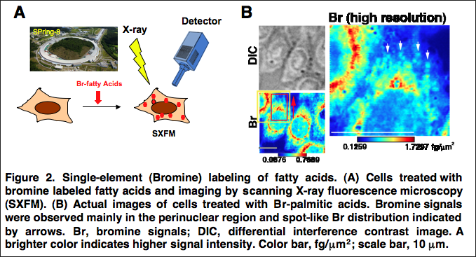
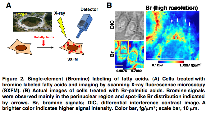
International researchers, including those from Osaka University, developed a new method to image intracellular fatty acids at a single cell level. They treated cells with fatty acids containing a single bromine atom and used scanning X-ray fluorescence microscopy to observe the molecules inside the cells. The technique offers superior resolution. The new method may improve understanding of the role of fatty acids in cell function and disease.
Fatty acids are essential for cell growth and survival, providing energy and forming important components of cell membranes. Because of their importance in cells, fatty acids' functions and metabolism, or breakdown have been widely studied by biologists, but the standard techniques makes it difficult to visualize precisely where these molecules are located within the cells.
A new method, recently published in the The FASEB Journal by an international team including Osaka University researchers, can now image intracellular fatty acids at a single cell level. The technique will allow researchers to understand the dynamic changes fatty acids undergo and how the cells use them. The method does not show cytotoxicity and offers higher resolution than standard imaging techniques.
"We use a technique called scanning X-ray fluorescence microscopy," study co-author Satoshi Matsuyama says. "This allows us to visualize trace elements inside cells, which means we don't need to label the molecules with chromophores to be able to see them."

Attaching optical fluorescent molecules, or chromophores, to biological substrates is common in cell-imaging studies, but these substituents are large and often interfere with cell metabolism, damaging the cells.
The researchers instead attached a single bromine atom to fatty acids, a process that required no complicated reaction steps, and treated cells with these modified fatty acids. The bromine was well known for bioimaging, and cells took up and metabolized the bromine-containing fatty acids..
Next, the researchers used their microscopy technique to find where in the cells the fatty acids and their metabolized derivatives were located. Their method was more sensitive and had a higher resolution than standard techniques, and they mainly detected the bromine-containing molecules in the perinuclear region and showing spot-like distribution.
"Fatty acids and their derivatives are important for cell function and are related to several diseases," co-author Kazuto Yamauchi says. "Knowing precisely where they are found in the cells will improve our understanding of their functions."
More information: M. Shimura et al. Imaging of intracellular fatty acids by scanning X-ray fluorescence microscopy, The FASEB Journal (2016). DOI: 10.1096/fj.201600569R
Journal information: FASEB Journal
Provided by Osaka University


