Inside the advanced photon source

To see inside Argonne's Advanced Photon Source (APS), all it takes is a little bit of light.
The electrons traveling around the kilometer-long electron storage ring generate extremely powerful X-rays that penetrate to the heart of some of our country's most complicated scientific questions.
Virtually every day, around the clock, scientists around the ring are simultaneously working on experiments in many different fields: materials science, protein biology, environmental science, chemistry, and nanotechnology, to name just a few.
This is possible at the APS because it simultaneously generates X-rays-at 35 different sectors, each containing one or more beamlines—which means that dozens of different experiments are going on at any given moment. Every year, thousands of users from industry, academia, and government laboratories come to Argonne to perform experiments at the facility.
The APS, one of the world's most powerful X-ray sources, produces pulses of light that are both extremely bright and penetrating. Together, these qualities let scientists look at all kinds of remarkable structures under different conditions, pressures, and temperatures.
Big Pharma, Tiny Protein
Several of the beamlines at the APS cater to users who come to Argonne from the pharmaceutical industry. Many new drugs that wind up on pharmacy shelves are inhibitors, which prevent the action of key cellular enzymes involved in certain diseases. The way that these enzymes work is by latching onto other molecules; if you can block the "active site" where that happens, the enzyme protein can't work.
To make a new drug, scientists build an inhibitor to precisely block that site—but they often have to be fashioned molecule-by-molecule, like making a key to fit into a lock. The first step, though, is knowing what the lock looks like.
To get a picture of the protein's structure, researchers bring tiny samples of crystallized protein to the APS, where they are loaded onto small pins and exposed to the APS X-rays. The patterns recorded by the beamline's detector can be interpreted to determine the positions of individual atoms within the sample. Research done at the APS has resulted in the development of drugs that treat HIV/AIDS (Kaletra) and melanoma (Zelboraf) and led to new insights into the structures of the viruses that cause HIV, influenza, swine flu, and even the common cold.
One of the sectors at the APS is owned and operated by Eli Lilly, the fifth biggest pharmaceutical company in the United States. Another, known as IMCA-CAT, is owned and operated by the Industrial Macromolecular Crystallography Association, a consortium including Merck, Pfizer, Bristol-Meyers Squibb, Novartis, and Abbott Laboratories.
Real Materials, Real Conditions
One of the major advantages of the X-rays produced by the APS is that they allow scientists to study materials in situ – that is, under a wide range of different environments. For example, researchers can examine how a catalyst will react at different temperatures or pH.
A little ways farther around the APS ring, Argonne chemist Peter Chupas and his colleagues look at the performance of battery components at the X-ray Science Division's Sector 11 beamlines.
"Years ago, we could only take one battery and cycle it very slowly, but now in the same amount of time we can investigate ten batteries," Chupas said. "By getting more information we can understand entire battery systems; we can get the real hard numbers on their performance so that we can go back to our models and improve our predictions about their behavior."
The X-ray beams at Sector 11 are both strong enough to penetrate all the way into a battery and narrow enough so that researchers can pick out specific regions to investigate. These narrow beams allow scientists to take data from many different points on the sample, creating a richer picture of a material's structure and function.
Like all of the APS beamlines, Sector 11 is used year-round by academic and industrial groups who come to the facility to try to bridge the gap between the fundamental structure and properties of materials and fully mature technologies. "What separates us from the pack is the fact that we're able to do these very precise and fundamental measurements on an applied system," Chupas said. "We're always looking for new ways to combine the two."
Argonne researchers are continually improving the instruments on the beamlines to provide users with the best technology available. "We're developing new tools constantly," Chupas said.
Honey, I Shrunk the Beam
The ability to condense beam sizes ever further has also yielded benefits at a life science beamline where researchers work on targeted drug design, especially for treating cancer. The development of the hard X-ray minibeam quad collimator, which earned an R&D 100 award in 2010, has let scientists shrink the beam diameter – from 20 microns down, in some cases, to a single micron. That's smaller than a strand of spider silk.
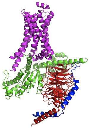
In one of the most challenging experiments at this beamline last year, researchers from Stanford University and other institutions wanted to study a type of protein called G-coupled protein receptors, a frequent drug target. Specifically, they wanted to study a protein that's part of an elusive but extremely important multiprotein complex.
The crystallized proteins are essentially invisible to the eye, which makes them difficult to position within the X-ray beam. The team had to create special computer software to scan the samples.
This beamline has also facilitated research on adenovirus, a virus of interest not only for its role in the common cold and other respiratory diseases, but also for its potential for gene-delivery methods for treating serious diseases like cancer.
Lighting up the future
Even though the APS already produces some of the brightest X-rays in the Western Hemisphere, Argonne engineers and physicists are already planning to make the facility even more powerful by dramatically improving the brightness, sensitivity, and resolution of the synchrotron.
A proposed upgrade to the APS, which received a preliminary go-ahead from Congress, has already entered the design phase. This year, the U.S. Department of Energy Office of Science, which funds the APS, allocated $20 million towards the upgrade. The APS Upgrade project plans to add more than a dozen new or upgraded beamlines and will dramatically improve X-ray beam properties, making the beam brighter at high energies and reducing the duration of the pulse.
"Each pulse can be thought of as a single exposure, like a snapshot," said Linda Young, director of the X-ray Sciences division at Argonne. "With shorter pulses, you can freeze the motion of faster processes— such as molecules reconfiguring under the direction of laser light."
Right now, the APS generates X-ray pulses that are approximately 100 picoseconds long—that's just one-tenth of a nanosecond—but researchers at the facility are trying to reduce that by a factor of nearly one hundred.
"We're just in the first phases of what we hope will result in the creation of a machine capable of exploring a wider range of materials and phenomena than ever before," said project director George Srajer.
In addition to the APS Upgrade, the APS will gain a brand new beamline—the only one of its kind—to explore shock physics: the branch of science that studies what happens to materials when they're exposed to large stresses in very short periods of time.
"The whole point of the upgrade is to provide more flexibility for our users so that they have access to state-of-the-art investigative tools, now and in the future," Srajer said.
Provided by Argonne National Laboratory


