This article has been reviewed according to Science X's editorial process and policies. Editors have highlighted the following attributes while ensuring the content's credibility:
fact-checked
trusted source
proofread
With living robots, scientists unlock cells' power to heal

Near the entrance to Michael Levin's lab at Tufts, four deer antlers are mounted on wooden boxes. They represent an incredible feat of regeneration in mammals: Deer shed their antlers annually and regrow the bone, blood vessels, nerves, and skin at a rate of half an inch per day.
Human regenerative abilities are much more limited. While we can grow scar tissue to heal wounds, knit fractured bone back together, and even regrow portions of some organs, this regeneration isn't as quick or complex as that of deer antlers—and we certainly can't regrow a lost leg the way some amphibians can. At least, not yet.
Levin, A92, Vannevar Bush Professor of Biology in the School of Arts and Sciences, is convinced that our cells have untapped regenerative capabilities, if only we can learn to speak their language. His lab is working to unlock the full potential of our cells, providing new ways to fight cancer, reverse degenerative diseases, repair congenital anomalies, and perhaps someday even regrow limbs.
Unlike other approaches to regenerative medicine, which may involve gene editing or stem cell therapy, Levin's goal is to take advantage of what the body already knows. The steps involved in creating an eye or a limb are too complex to micromanage, he said. But perhaps—with the right set of signals—we can give the body a new goal and let intelligent groups of cells manage the details of how to achieve it.
"We don't want to try to tell every cell and every gene what to do," Levin said. "We're not looking to teach cells how to grow a leg; we're looking to convince them that that's what they should do."
When Levin talks about the intelligence of cells, he doesn't mention brains. He cites a definition from William James, an American psychologist and philosopher in the late 19th century, who described intelligence as the ability to achieve the same goal in multiple ways.
By this definition, a system is intelligent if it can have and achieve goals, even when faced with unexpected hurdles. The system could be a creature, a machine, an organ, or a single cell; if it can problem-solve, it has a kind of intelligence.
Individual cells can solve simple problems and have simple goals—survival and reproduction. But once cells start to work together, they create a collective intelligence capable of remembering and achieving larger goals, such as forming the systems and organs that make up a body.
It's this collective intelligence that Levin hopes to understand and harness. Even as new discoveries in the lab create opportunities for developments in biomedicine or other areas, they provide tantalizing pieces of information about how cells talk to each other and what they are capable of. It's a puzzle that Levin has been putting together for most of his career, and one that he expects will take at least another decade to complete.
"Our work is not solved," Levin said. "We know cells are talking to each other, but we don't know what they're saying yet. We still need to crack that code."
The cognitive glue for cells
Cells in a developing embryo need to be in constant communication, working together to determine what to build and where to build it. They store and send these communal blueprints in electrical signals, indicating left and right sides, which tissue should become organs, or when to stop building toes.
With the correct tools, Levin and his colleagues have been able to view these signals, watching tadpoles develop an "electric face"—patterns of activity that show the future locations of the eyes, nose, and mouth—long before the cells started to form those structures.
"Bioelectricity allows a bunch of cells to connect into a network that can compute and store memories of much larger goals," Levin said. "Your individual cells have no idea what a finger is or how many fingers you should have, but a collective of cells absolutely does."
Cells create and share bioelectric signals through gateways in the cell membrane called ion channels. Ion channels allow certain charged molecules to pass through the membrane, creating different charges inside and outside of the cell, akin to the positive and negative sides of a battery. The movement of ions through the membrane creates tiny electric currents.
Levin describes bioelectricity as "the cognitive glue holding our cells together." In the brain, where groups of neurons rapidly shuffle ions around to create strong, fast signals, bioelectricity controls how we process information and move through space. In other parts of our bodies, where the signals are smaller, slower, and often overlooked, it holds a sort of collective memory that tells cells how to create the physical structures our body requires and how to respond when those structures are damaged.
By learning to decipher and manipulate these subtler bioelectric signals, Levin hopes to find new ways to help our body heal itself.
The lab has made a variety of promising discoveries that reveal some of bioelectricity's therapeutic potential.
In one experiment, the researchers were able to use a computational model to predict the normal voltage patterns for a developing frog embryo and determine how those patterns were being disrupted by exposure to nicotine, which causes abnormal brain development. The researchers found a treatment that would restore normal voltage patterns. As a result, frog embryos were able to repair and recover from nicotine-induced defects.
Levin's team has also been exploring a bioelectric approach to cancer treatments. Viewed through the lens of bioelectricity, cancerous cells are ones that have become disconnected from the cellular communication network and are acting as individuals. Levin and his colleagues have shown that blocking some ion channels and restoring normal bioelectric patterns can reconnect cancer cells to the larger network and rein in the rogue cells. Despite cancerous mutations, the cells behave normally.
"They do the right thing because they're sharing all their memories with other cells through these electrical synapses, and their goals are now organ-level goals," Levin said. "They no longer just want to make copies of themselves."
The researchers also determined that they could identify early stages of cancer formation in tadpoles by tracking unusual variations in the electrical patterns of individual cells. The discovery could spur the development of new bioelectric diagnostic tools.
The team has even made progress toward the goal of regenerating whole body parts. Levin and David Kaplan, Stern Family Endowed Professor of Engineering, created a device packed with a cocktail of drugs intended to tweak the behavior of cellular ion channels and encourage growth. The device allowed an adult frog—which can't usually regenerate limbs—to regrow a functional leg.
Impressively, the device needed to be applied for only 24 hours to jump-start the regeneration process. The two researchers have created a company called Morphoceuticals to develop this work for clinical applications, starting with studying its use in mammals.
"We aren't using any external stimulation; we are using the same interface that cells and tissues use to hack each other in the body," Levin said. "By tweaking these ion channels, we can play that interface like a piano and get it to have different kinds of electrical computations."
The challenge is in determining what tune the researchers need to play to get the behaviors they're looking for.
In tadpoles, Levin and his colleagues can convince groups of cells to create functional eyes, hearts, and limbs. In planaria—small, brown flatworms with a remarkable capacity for regeneration—Levin's team can encourage the growth of multiple heads and even cause one species to develop the head of a separate, closely related species, revealing the kinds of information different signals can carry.
In other organisms, the researchers are examining voltage patterns associated with aging and trying to reverse those effects. But the team doesn't yet have a cohesive understanding of how to interpret the specific signals that cells are sending and how to persuade the cells to, say, repair damaged organs or fend off a degenerative disease.
"At the moment, we're taking all these independent phenomena that are linked by the same process and trying to develop a deeper understanding of the biology of it," said Patrick McMillen, a staff scientist who has worked in Levin's lab for the last seven years. Each experiment reveals more about the previously overlooked role of bioelectricity and brings the researchers closer to being able to unlock the same potential in human cells.
Learning to decode bioelectric signals
For many years, researchers studying bioelectric communication in the human body focused on our brains. Neural communication is fast and strong: Neurons can fire 80 millivolt signals in a matter of milliseconds.
By contrast, bioelectric signals in the rest of the body are closer to 3 millivolts and can take minutes or hours to develop. Established techniques for making images of bioelectricity in neurons can't effectively detect bioelectricity in other cells. So the importance of bioelectric communication outside the brain was largely overlooked until recently.
"The things we're looking for are very, very subtle and very, very slow," said Patrick McMillen, a staff scientist in the Levin Lab. "We've been working for a long time to find tools that allow us to see these sorts of signals."
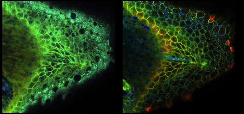
The researchers use a technique called fluorescent lifetime imaging microscopy, staining cells with a fluorescent dye that glows an array of different colors in reaction to the presence of various molecules that affect the cells' bioelectric state. The resulting rainbow images allow the researchers to detect and measure subtle physiological changes, which indicate changes in the bioelectricity. McMillen recently developed a new strategy for using these techniques in live frog embryos.
"I'm deeply optimistic that this approach will enable us to read the bioelectric code in developing animals and complex tissues with unprecedented clarity and precision," McMillen said.
The ability to image neurons as they fire has helped us understand how our brains work and how to treat the diseases that affect it. The researchers hope that being able to detect the subtler bioelectric signals underpinning cellular communication in the rest of our body will open the door to similarly revolutionary advancements in biomedicine.
Biobots and beyond
Levin's work continually shows that collectives of cells are capable of much more than we typically give them credit for. They are problem-solving machines, and, when given new instructions, they can accomplish tasks far beyond what they normally do in the body.
The clearest example of this is the lab's biobots, microscopic "robots" made of living cells that have been cut loose from an organism's bioelectric signals to become independent actors. They have been "freed from the constraints of being part of a body and allowed to be whatever they can be," as Levin put it, and this freedom has allowed them to develop incredible and unexpected abilities.
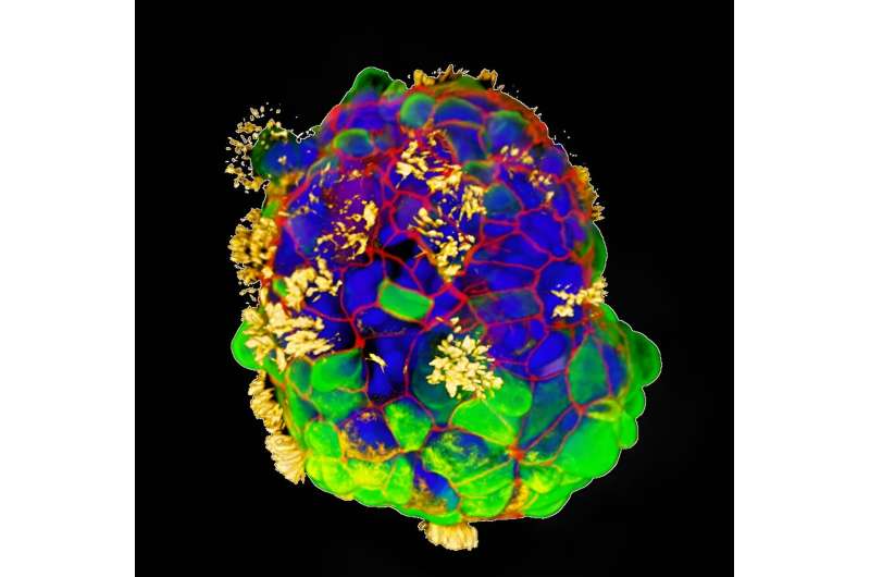
The lab's Xenobots are millimeter-wide blobs made from several different kinds of cells taken from Xenopus laevis, the African clawed frog. The Xenobots live for about a week and can repair themselves when damaged. The researchers used a combination of muscle cells and skin cells with hairlike projections called cilia, which would typically move mucus across the frog's skin, to design the Xenobots to propel themselves across a petri dish and work together to sweep specks of debris into a pile.
"We've learned a lot of new rules about how cells self-assemble in tissues that are not obvious when looking at an intact system," said Doug Blackiston, the senior scientist in the Allen Discovery Center who sculpted the original Xenobots by hand. "By moving things around and playing with the biology, you can get new insight into the basic science."

Blackiston sees possibilities for using the Xenobots in the environment. The tiny clumps of cells could carry sensors to measure environmental pollutants, such as BPA (a chemical used in some plastics), or drug compounds in wastewater. Or they might be used to collect and concentrate rare metals for easier extraction. They are also a steppingstone to other discoveries. In November, the lab announced the creation of Anthrobots—similarly active clusters grown from adult human tracheal cells.
The researchers used tracheal cells because they have cilia, akin to those of a frog's skin cells, making it easier for the Anthrobots to develop the ability to move. And like the Xenobots, the Anthrobot cells exhibit unexpected behaviors beyond what they would do if they were a part of the body. When the researchers added a cluster of Anthrobots to a damaged sheet of nerve cells, for example, the Anthrobots parked themselves across the damaged area, and the nerves beneath them began to heal.
"These bots can move across a damaged site, sit there, and actually help the nerves knit across, essentially repairing the damage in the course of three days," said Gizem Gumuskaya, AG23, who is the lead author on a recent paper and conducted the work as part of her doctoral thesis in Levin's lab. "Could we take cells from a patient, make personal Anthrobots, and use them to help heal neural damage? That's the application we're working toward."
Because the Anthrobots would be created from a patient's own cells, they would be able to move through the body without being attacked by the immune system. And healing neural damage is just the start of what they could enable. Levin imagines Anthrobots dropping off regenerative molecules, chasing down cancer cells, cleaning the plaque from arteries, and who knows what else.
"We've already shown they can heal neural wounds, and that's just the baseline. We haven't even started programming them yet," Levin said. "They are an amazing platform for biomedicine, and also a sandbox in which we get to understand the collective intelligence of cells."
Provided by Tufts University


