This article has been reviewed according to Science X's editorial process and policies. Editors have highlighted the following attributes while ensuring the content's credibility:
fact-checked
trusted source
proofread
Elucidating the process of bile duct formation in the liver

Bile ducts are pathways that carry hepatocyte-produced bile from the liver to the small intestine. In the human fetal liver, bile ducts are formed from bile duct epithelial cells surrounding the portal vein, and hepatocytes form on the outside. Both bile duct epithelial cells and hepatocytes are formed by the differentiation of common progenitor cells (hepatoblasts).
Molecules (ligands) from portal vein cells act on receptors called Notch on the plasma membrane of hepatoblasts, which differentiate into bile duct epithelial cells (called the Notch signaling pathway). However, the molecular mechanism by which bile duct epithelial cells differentiate only along the portal vein remains unknown.
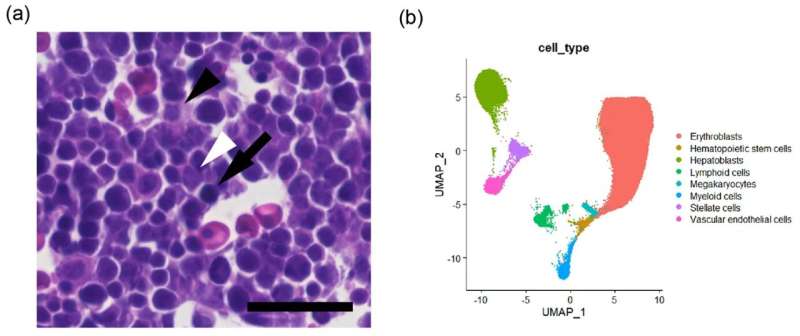
To understand this mechanism, researchers analyzed public data on the human fetal liver. They identified three major cell types in the human fetal liver: portal vein, hepatoblasts, and hematopoietic cells, which mainly become erythrocytes. Adult erythrocytes are produced in the bone marrow, whereas fetal erythrocytes are generated in the liver.
The study is published in BMC Research Notes.
In examining the ability of these cells to act as components of the Notch signaling pathway, researchers found that only portal vein cells were able to express the major ligand JAG1 and to be the sender of signals. Additionally, only hepatoblasts were able to express the major receptors, NOTCH1 and NOTCH2 and to be the recipients of signals. Hematopoietic cells do not possess this ability.
These results indicate that hepatoblasts along the portal vein receive Notch signaling and differentiate into bile duct epithelial cells, whereas, at a distance from the portal vein, hematopoietic cells and hepatoblasts act as barriers and do not transmit signals, resulting in the failure of differentiation into bile duct epithelial cells.
The research team has been using mathematical analysis to verify the conditions under which Notch signals propagate widely or are localized in tissues. These findings are consistent with the conditions for localized Notch signaling (slow production of ligand or receptor) obtained in the mathematical analysis and provide molecular biological support for the previous mathematical analysis results.
The elucidation of the molecular mechanism of bile duct production in the liver is important for the future development of artificial organs.
More information: Masaharu Yoshihara et al, Chromatin accessibility analysis suggested vascular induction of the biliary epithelium via the Notch signaling pathway in the human liver, BMC Research Notes (2023). DOI: 10.1186/s13104-023-06674-8
Provided by University of Tsukuba


