This article has been reviewed according to Science X's editorial process and policies. Editors have highlighted the following attributes while ensuring the content's credibility:
fact-checked
peer-reviewed publication
proofread
Realizing in situ electron paramagnetic resonance spectroscopy using single nanodiamond sensors

Researchers have used the nitrogen-vacancy (NV) center inside a single nanodiamond for quantum sensing to overcome the problem of random particle rotation. Their study is published in Nature Communications.
Being able to detect and analyze molecules under physiological in situ conditions is an important goal in the field of life sciences. Only by observing biomolecules under this condition can we reveal conformation changes when they realize physiological functions.
Thanks to its high sensitivity, good biocompatibility, and the characteristics of magnetic resonance detection of single molecules at room temperature atmosphere, the NV center quantum sensor is more suitable for physiological in situ detection than traditional magnetic spectrum resonance instruments.
However, the results of tracking the movement of nanodiamond in living cells show that it rotates randomly both inside the cell and on the cell membrane, making the current common magnetic resonance detection methods ineffective.
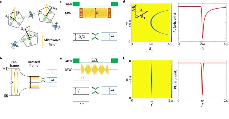
To solve this problem, the research team designed an amplitude-modulation sequence, which will generate a series of equally spaced energy levels on the NV center.
When the energy level of the NV center matches the energy level of the measured target, resonance will occur and the state of the NV center will change.
By scanning the modulation frequency, the electron paramagnetic resonance (EPR) spectroscopy of the target can be obtained, and the position of the spectral peak is no longer affected by the spatial orientation of the NV center.
In this work, the ions in the solution environment of nanodiamond were measured by EPR spectroscopy under the condition of in situ. The research team simulated the movement of nanodiamonds in the cell to detect the solution of oxygen vanadium ions.
When there is nanodiamond rotation, it is difficult to conduct accurate quantum manipulation of NV centers, but zero-field EPR spectrum of oxo-vanadium ions can still be measured.
This result proves in principle that it is feasible to use the NV center in nanodiamond to realize the detection of intracellular physiological in-situ magnetic resonance.
The oxygen vanadium ions detected in this work itself have biological functions. The ultra-fine constant of oxygen vanadium ions can be analyzed and obtained by the EPR spectrum measured by a single moving nanodiamond.
The research team has previously relaxed the detection conditions of single-molecular magnetic resonance detection from solid conditions to an aqueous solution environment, and this work has further promoted it to the in situ environment.
The researchers were led by Prof. Du Jiangfeng, Prof. Shi Fazhan and Prof. Kong Fei from the University of Science and Technology of China of the Chinese Academy of Sciences.
More information: Zhuoyang Qin et al, In situ electron paramagnetic resonance spectroscopy using single nanodiamond sensors, Nature Communications (2023). DOI: 10.1038/s41467-023-41903-5
Journal information: Nature Communications
Provided by University of Science and Technology of China


