This article has been reviewed according to Science X's editorial process and policies. Editors have highlighted the following attributes while ensuring the content's credibility:
fact-checked
trusted source
proofread
Mix of X-ray methods offers improved structure analysis

Sometimes scientists have to accept that a method they have used for years fails under certain conditions. Such a failure calls for a careful analysis of the shortcomings and their subsequent elimination. An international team from the University of Regensburg, the University of Durham and the Helmholtz-Zentrum Dresden-Rossendorf (HZDR) has now done precisely this.
They effectively reinvented X-ray diffraction, a widely used method for determining crystal structures, by combining it with X-ray spectroscopy, thereby removing one of the crucial drawbacks of the method, known for over a century. One of the researchers has now received the Lieselotte Templeton Prize of the German Crystallographic Society for this achievement.
X-rays are not only useful imaging the human body but are also valuable for examining novel materials: they can be used to find out how crystals are structured in detail—indispensable information, for example, for the development of new high-tech materials or drugs. This method, known as X-ray diffraction, has been around for a long time, but some time ago, a team of researchers came across a fundamental methodological problem.
Dr. Michael Bodensteiner's group at the University of Regensburg examined a copper compound with X-rays at a rather unusual "X-ray color," the so-called K? radiation. "We used an almost perfect crystal and actually expected to be able to determine its structure precisely," the chemist explains. "But then we found that at some point something physically nonsensical came out. Simply put, the copper atoms were not sitting in the crystal lattice as they should have been."
To get to the bottom of the mystery, the team took a closer look at the experimental method. In doing so, the researchers discovered that particular corrections involved in the process were distorting the results rather than improving them. "In the past, these mathematical procedures were usually quite sufficient," Dr. Bodensteiner explains. "Now, our instruments provide data of such precision that these corrections reach their limits and therefore have to be improved."
Breakthrough in Grenoble
To overcome this limitation, the Regensburg scientists teamed up with HZDR researcher Dr. Christoph Hennig. He works in a rather special place—the European Synchrotron (ESRF) in Grenoble, France. Compared to the conventional X-ray tubes of a university lab, the accelerator-based facility delivers a much more intense and highly focused X-ray beam. In Grenoble, the HZDR operates an experimental facility, the Rossendorf Beamline (ROBL).
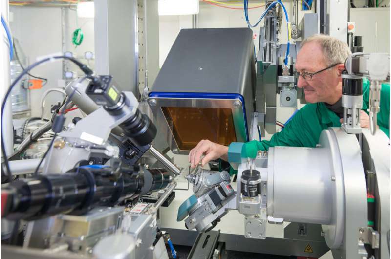
"It provides very good conditions for such measurements," says Dr. Hennig. "Among other things, there is a powerful diffractometer that can take high-resolution diffraction images," that is, it can measure very precisely how X-ray radiation is affected by passing through a crystal structure. Simultaneous spectroscopic measurements are also possible—a designated specialty of ROBL. In this process, a sample is illuminated with alternating "X-ray colors." This approach allows conclusions to be drawn, for example, about the chemical properties of the elements that constitute a crystal.
The team's idea was to combine both methods, X-ray diffraction and spectroscopy—an approach that has rarely been tried before. "One of the challenges was to coordinate the various components of the equipment, such as the detectors that capture the scattering intensities," says junior researcher Florian Meurer. In their experiments, the researchers then targeted those measurement points primarily where the conventional method yielded unreliable results. "By combining X-ray diffraction and spectroscopy, we came up with consistent values. This clearly proves that our method works." For his master's thesis on this project, he will be awarded the Lieselotte Templeton Prize of the German Crystallographic Society.
Perspectives for deep geological repository research
Although the researchers still need to refine their method, it holds great promise for the future. For example, it should be possible to analyze the structures of certain crystals more precisely than before.
"In addition to pure structural information, we should also be able to learn more in one and the same measurement, for example about the oxidation state of an element," says Dr. Michael Bodensteiner. "That would be helpful, for example, for studying catalytic reactions in chemistry." The combined method of X-ray structure and spectroscopy should also be useful for future projects at the ROBL in Grenoble. Dr. Hennig and his team are studying the behavior of radioactive substances such as those present in nuclear waste.
"We hope to be able to determine the structures of certain radioactive molecular compounds more precisely," says HZDR crystallographer Dr. Christoph Hennig. "This would allow us to better assess whether a particular substance will remain permanently in a repository or whether it may eventually be released into the environment."
More information: Florian Meurer et al, Refinement of anomalous dispersion correction parameters in single-crystal structure determinations, IUCrJ (2022). DOI: 10.1107/S2052252522006844
Provided by University of Regensburg


