February 2, 2022 feature
A device based on 3D transistor arrays for collecting intra and inter-cellular recordings

Animal cells can use elements or ions to generate electrical impulses. These impulses are then conveyed from one cell to another, traveling across cellular networks.
The ability to precisely record electrical signals exchanged by cells could aid research and improve practices in numerous health-related fields, including cardiology and neurology. Most existing technologies, however, are limited in both their sensing accuracy and scalability.
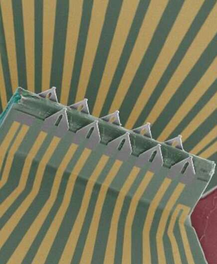
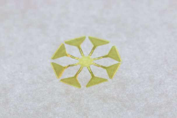
Researchers at the University of California San Diego have recently developed a highly sensitive sensing device that could be used to record the electrical signals of cells with greater precision. This device, introduced in a paper published in Nature Nanotechnology, is comprised of multiple sensors, which can collectively measure the propagation of electrical signals exchanged by different cells or inside individual ones.
The recent study was led by Dr. Yue Gu while working in Prof. Sheng Xu's lab at UC San Diego. Dr. Gu, is now a postdoctoral associate at Yale University.
"The establishment of our 3D structure, also known as a 'pop-up' architecture, is based on a unique method, the compressive buckling technique that I developed during my postdoctoral studies in 2015," Prof. Xu, one of the authors of the recent paper, told Phys.org. "The compressive buckling technique takes advantage of conventional and versatile cleanroom microfabrication techniques to generate sophisticated 3D structures."
The 3D 'pop-up' structures used by Prof. Xu and his colleagues can be built using a wide range of materials that are compatible with microfabrication techniques. The materials they are made of can in turn determine their function, which could be electromagnetic wave attenuation, mechanical vibration, pressure, and strain sensing, or electrical signal sensing.

In their study, the researchers set out to build these 3D structures so that they could be used to precisely record electrical signals generated and exchanged by cells. Their key objective was to effectively leverage the versatility of the compressive buckling technique to build a device that would collect accurate intra- and inter-cellular recordings.
"Embedding semiconductive materials and engineering transistors in this pop-up architecture extends the application of the technique," Prof. Xu explained. "Our determination to apply this structure to cells, specifically cardiac muscle cells, was sparked by discussions that Dr. Gu and I had with cardiologists and neurologists back in 2015, who were complaining about the difficulties of recording intracellular signals using the existing tools, such as the patch-clamp, which is the gold-standard for recording cellular electrical signals."
After they learned about the challenges that medical researchers were experiencing when trying to collect precise recordings of cellular electrical signals, Dr. Xu and Dr. Gu started experimenting with unique engineering approaches that could simplify their work. Ultimately, this led to the development of the new sensor array introduced in their recent paper.
"Another objective of our study was the implementation of intracellular sensors into 3D engineered cardiac tissues," Prof. Xu said. "It is well known that the electrophysiological properties of cells vary when they are in live animals, isolated from the live animals and cultured in dishes. Recording the signals in-vivo is always the most significant and yet challenging step."
Prof. Xu and his colleagues were the first to collect precise intracellular recordings of cells within the engineered cardiac tissue. Their study could thus be a first step towards the collection of reliable, in-vivo cellular recordings.
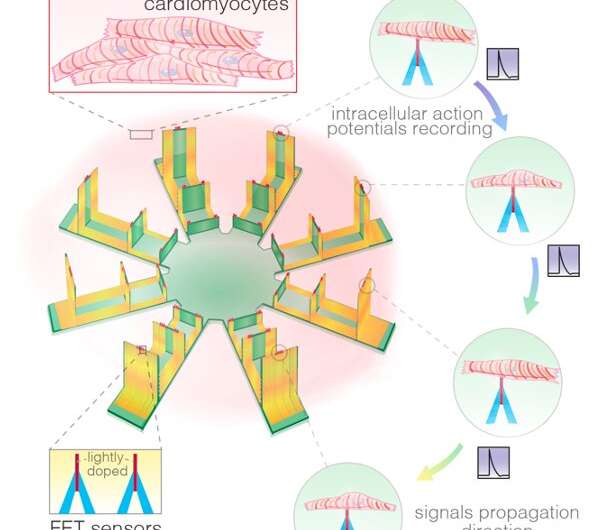
"Cell membrane potential biasing the gate terminal of individual transistors results in a change in the current from the drain to the source terminal of the transistors," Prof. Xu explained. "Therefore, current fluctuations reflect the momentary membrane potentials. The multiple transistors in the array we developed can simultaneously record signals from different positions of a cell or different cells."

To monitor signal propagation behaviors inside and between cells, the researchers' device sequences the signals picked up by its many transistors. In contrast with other previously proposed methods for collecting cellular recordings, the new device is capable of monitoring multiple cells simultaneously. In addition, its transistors can retain intact full-amplitude cell membrane potentials, without suffering from attenuations or impedances associated with the process through which it accesses cells.
"Functionalized transistor surfaces by phospholipid bilayer materials can also camouflage the inorganic transistors to be cells, which greatly facilitate their insertion into the cell body," Prof. Xu explained. "In such conditions, internalization is described as a spontaneous fusion process, which leaves minimal even no invasiveness to the cell."
The sensing device developed by Prof. Xu and his colleagues can also monitor the electrical signal conduction velocity inside a cardiomyocyte. This measurement can be of vital importance for the work of cardiologists, as comparing it to the conduction velocity between neighboring cells can aid the detection and understanding of some cardiac diseases, including cardiac fibrosis.
"As part of our study, we deployed the transistor array into 3D cardiac tissue and recorded the intracellular electrical signals of single cells for the first time," Xu said. "In the process, we also recorded the conduction of electrical signals and calculated their velocity."
So far, the researchers have primarily tested their transistor-based sensing device on cardiac tissue, attaining highly promising results. Their initial findings suggest that it could eventually be used to gather precise recordings of electrical signals produced and exchanged by cells, both in laboratory settings and in-vivo, on the brains or hearts of live animals or human patients.
"We are now pursuing several new goals," Xu added. "The first is to use our transistors to perform in-vivo tests on intact hearts or brains. The second is recording the intracellular activities of neurons at different neuronal locations. Finally, as some endocrine cells are also electrogenic, meaning that their electrical activities are related to other physiological events, they are also of great interest."
More information: Three-dimensional transistor arrays for intra- and inter-cellular recording. Nature Nanotechnology(2021). DOI: 10.1038/s41565-021-01040-w.
Journal information: Nature Nanotechnology
© 2022 Science X Network


