Scientists produce the first anatomical atlas of Asian citrus pest

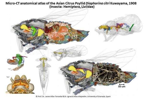
Scientists from the University of Granada (UGR) have made the first anatomical atlas of a tiny insect measuring just 3 mm called Diaphorina citri, the Asian citrus psyllid (ACP), which responsible for the greatest economic losses in citrus crops worldwide.
As a pest, Diaphorina citri is very harmful to citrus trees (lemons, limes, oranges, grapefruits, tangerines, and kumquats ) due to the bacteria it spreads (Candidatus Liberibacter spp.). These bacteria cause Huanglongbing (HLB) disease (also known as citrus greening disease), considered the most serious bacterial disease affecting citrus fruits, as the bacterial infection causes yield losses, small, bitter, and foul-tasting fruits, and, finally the death of the tree. This translates into annual losses of millions of dollars.
To date, this species has not been detected in the Iberian Peninsula, although another related species (Trioza erytreae) capable of transmitting the bacteria that cause Huanglongbing has already spread. Diaphorina citri could potentially cause devastating damage to the economy of those Spanish regions with large-scale citrus production, and the relevant authorities must make every effort to avoid the introduction of this insect (or diseased plants) when importing citrus fruits from countries where the disease and this vector species are present.
The research group, led by Professor Susan J. Brown from Kansas State University, conducted a multidisciplinary macro-project to study the insect, the bacteria it transmits, its effects, and means of control (citrusgreening.org).
The US research team approached Javier Alba-Tercedor of the UGR's Department of Zoology to lead the study of the functional anatomy of the insect using microtomographic techniques. Alba-Tercedor has an extensive track-record in the microtomographic study of insects, and his work (including photos and videos) has received various international awards. Given their spectacular aesthetic qualities, these have also been widely disseminated by the media.
Following a formal agreement between the UGR and Kansas State University, including collaboration with Dr. Wayne Hunter (from the Department of Agriculture in Fort Pierce, Florida), the insects were obtained for the study.
This study forms part of the doctoral thesis of Ignacio Alba-Alejandre (supervised by Alba-Tercedor). A recent article published in the prestigious journal Scientific Reports (by Nature) closes the series of articles providing a highly detailed vision of both the external and the internal structures of this insect.
This study offers the first complete micro-CT reconstruction of this pest and constitutes a ground-breaking detailed anatomical study of a psyllid as the first anatomical micro-CT study of a hemipteran to be studied in its entirety. And, together with other papers already published by this team from the UGR on the coffee borer beetle, these studies represent the anatomical reconstructions of the smallest insects carried out to date using micro-CT.
Based on the descriptive aspects of the study, the team was able to elucidate the operation of different structures. Among the most noteworthy examples are the male sperm pump—an impellent suction pump that serves to expel sperm—and, similarly, the spermatheca (sac) in which females store sperm. This, too, acts as an impellent suction pump, performing an inward sucking motion, retaining the sperm after copulation, storing and nourishing them, and later, at the point of laying, contracting the walls of the sac to expel the sperm and fertilize the eggs. This mechanism requires a complex series of special valves and muscles.
An articulated reproductive organ
The complexity of the articulated reproductive organ of males is particularly striking, as is the fact that females have a more voluminous nervous system than males. The latter phenomenon undoubtedly occurs because they have to perform more complex vital functions, such as selecting the ideal location for subsequent egg-laying.
Curiously, the hindgut of the female is posteriorly differentiated into a rectum that forms a small rectal ampulla, into which it deposits small amounts of excrement. By contracting the walls of the ampulla, the feces are violently propelled away from the body, thereby avoiding contact that could contaminate the eggs.
Another interesting discovery to emerge from the analysis carried out at the UGR was that these insects have glands at the base of the legs (coxal glands) and others at the base of the antennae (antennal glands) that produce sex pheromones. Thus, when the insect settles on the leaves, it impregnates them with the secretions of the coxal glands, which also attracts the opposite sex. Once males and females are close to one another, they begin a kind of "dance" in which they touch antennae and, thanks to the secretions of the antennal glands, recognize each other's sexes.
These are just a few examples of the many discoveries made during the course of the UGR study. In the words of the researchers, "we often feel like true explorers discovering new territories, contemplating structures from never-before-seen perspectives, uncovering previously unknown structures, and deducing the how and why of their function."
Scientific and aesthetic value
The work has produced numerous plates of figures that, as well as being of great scientific interest, have undoubted aesthetic value. Furthermore, the research team has been able to reconstruct in 3D—for the first time—an adult feeding on the leaf of a citrus fruit, showing how its stylets pierce the walls of the leaf to reach the vessels of the phloem and feed by sucking the sap. They have also produced a 3D model version for mobile devices (tablets and smartphones).
All of this work combined, together with numerous animated videos that enable viewers to see the structures in detail from multiple perspectives, renders this research a unique and extremely useful anatomical atlas not only for the study of this particular species but also for the investigation of insects in general. In addition, its impressive aesthetics make this material ideal for teaching and scientific dissemination purposes.
More information: Javier Alba-Tercedor et al. Using micro-computed tomography to reveal the anatomy of adult Diaphorina citri Kuwayama (Insecta: Hemiptera, Liviidae) and how it pierces and feeds within a citrus leaf, Scientific Reports (2021). DOI: 10.1038/s41598-020-80404-z
Journal information: Scientific Reports , Nature
Provided by University of Granada


