November 25, 2020 report
X-ray diffraction reveals details inside mummies without having to open them up

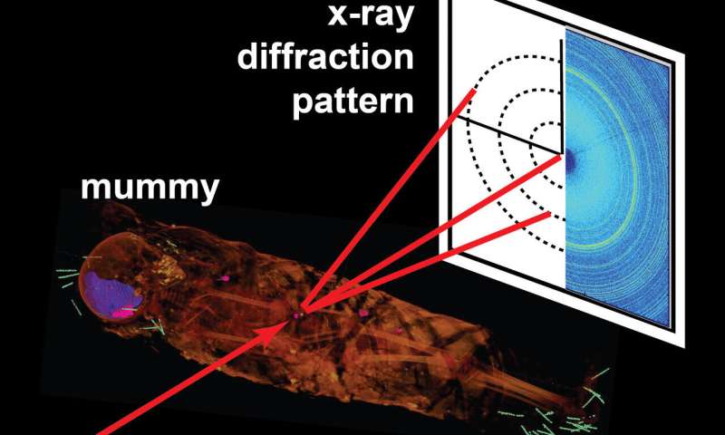
A trio of researchers from Northwestern University, Metropolitan State University of Denver and Argonne National Laboratory has found that using X-ray diffraction on mummies makes it possible to see inside the wrappings without opening them. In their paper published in Journal of the Royal Society Interface, S. Stock, M. Stock and J. Almer describe scanning an Egyptian mummy in two ways and what they found by doing so.
Egyptian mummies have captivated archeologists and historians alike for centuries. In the early years, the only way to know what was under the wrappings was to open the mummy and take a peek inside. In more recent times, X-ray technology has been used to visualize mummies without removing wrappings. In this new effort, the researchers have improved on this idea by using two different kinds of technology to get much higher resolution imagery.
The work involved first scanning a mummy called Hawara Portrait Mummy No. 4 with a computed X-ray tomography (CT) scanner to get images of the entire body inside the wrappings. They then examined the images produced by the CT scanner to find areas of interest. Then they used X-ray diffraction on those areas of interest to get a much more detailed look.
The researchers discovered one surprising fact: The person inside the wrappings was not an adult woman, as had been assumed, but was instead a five-year-old girl who had not yet developed her permanent teeth. Prior research had found that the mummy was from the first century, a time during which portraits were displayed on the outside surfaces of mummies, generally of the person preserved. Hawara No. 4 had a portrait of a grown woman. It is not known if the portrait was of someone else, or if the artist was imagining the girl as an adult.
The researchers also found multiple needle-like structures inside the mummy, which they assumed had been placed there by modern workers trying to preserve it. More interestingly, they found an object that had been placed over the abdomen—it measured just 7 mm long, was elliptically shaped and was made of calcite. The researchers assume that it was likely an amulet placed to conceal a wound or disfigurement.
More information: S. R. Stock et al. Combined computed tomography and position-resolved X-ray diffraction of an intact Roman-era Egyptian portrait mummy, Journal of The Royal Society Interface (2020). DOI: 10.1098/rsif.2020.0686
Journal information: Journal of the Royal Society Interface
© 2020 Science X Network


