Electron microscopy provides clues into the colorful chemistry of dragonfly wings

Dazzling dragonfly wings may send poets rhapsodizing, but scientists yearn for a better understanding. In particular, they want to know the chemistry of the different layers giving rise to natural photonic crystals that help create color.
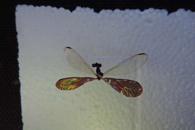
Now, a collaboration of Brazilian researchers from the Federal University of Minas Gerais, Belo Horizonte, Brazil, have teamed up with Minnesota experts in chemical surface imaging at Physical Electronics, Inc. (PHI) to puzzle out the color mechanism of the male Amazonian glitterwing dragonfly (Chalcopteryx rutilans).
Investigators will present their molecular surface imaging results and analyses during the AVS 64th International Symposium and Exhibition Oct. 29-Nov. 3, 2017, in Tampa, Florida. They analyzed both transparent and colored wings to correlate with the electron microscopy and optical results.
The colors of the glitterwing span the visible spectrum with shimmering red, blue, and yellow/green regions on the wings, the source of which they hope to find.
Brazilian investigators derived partial answers to this question using electron microscopy methods of scanning electron microscopy (SEM) and transmission electron microscopy (TEM). Probing the glitterwing color mechanics revealed that the iridescent wings have multiple alternating layers with different electronic densities. The variation of local color was related to the number and thickness of the layers, which changed across the wing.
While measurement of the thickness and number of layers was readily achievable by electron microscopy, the approach was unable to characterize the chemistry of the different layers giving rise to these natural photonic crystals. To fully understand the color mechanism, they needed to measure chemical structures in the wing.
By teaming up with Minnesota colleagues at PHI, they measured the actual chemistry in the wing structure with an advanced molecular surface imaging technique known as Time-of-Flight Secondary Ion Mass Spectrometry (TOF-SIMS). This extremely sensitive surface analytical technique can reveal highly detailed molecular and elemental data about surfaces, thin layers and interfaces in both 2-D and 3-D. TOF-SIMS can be used to probe the 3-D structure and chemistry of a wide variety of organic and inorganic materials, both synthetic and naturally occurring.
Among the most interesting findings the team discovered is that the periodic changes in local electron densities may correspond to variations in the sodium (Na) and potassium (K) concentrations through the thickness of the wing. They could not find any similar findings in the literature, however.
David M. Carr, an engineer and senior scientist at PHI, brings to light the importance of nature's engineering and its applications in technology development.
"Nature can often provide examples for engineering solutions. The whole field of biomimicry is devoted to learning from nature for potential solutions to difficult engineering problems," Carr said. "Every natural sample has unique features and a lot to teach us."
Provided by Science and Technology of Materials, Interfaces, and Processing


