April 27, 2017 report
Ultra-high resolution images of butterfly wing crystals offer clues to how nano-scale structures form

(Phys.org)—An international team of researchers has taken another step toward understanding the process by which butterfly wing scales develop crystals that result in bright, vivid colors. In their paper published in the open-access site Science Advances, the group describes the methods they used to study the wing scale crystals of the hairstreak butterfly—a black and bright blue native of parts of Colorado and northern Mexico—and what they found.
People of all backgrounds are captivated by butterflies—their bright colors and lackadaisical lifestyle of flitting from flower to flower tend to elicit smiles and warm feelings. And while a general understanding of the physical structure of the butterfly's wings has been well documented, the process by which they arrive at their coloring has never been discovered—this is because it happens over the course of several days inside of their cocoons, where tiny cameras would not really work. In this new effort, the researchers report taking another step in the discovery process.
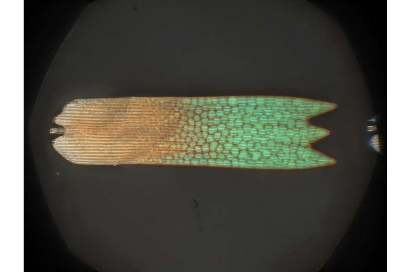
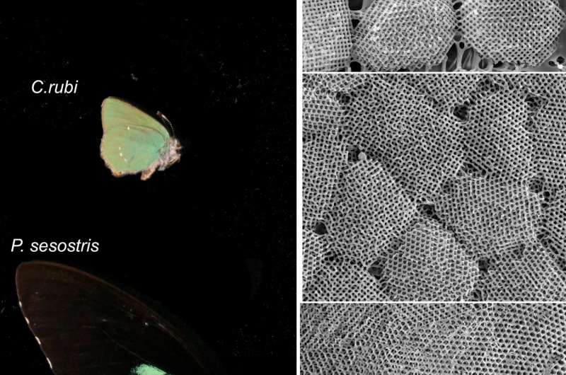
Prior research has shown that butterfly wings are covered in scales with chitin crystals called gyroids on their surfaces—the gyroids reflect light in certain ways, creating the perception of colors. But how the gyroids develop to display colors is still unclear. In this new effort, the researchers took the closest ever look at the scales and gyroids using several imaging techniques, and report finding something new.
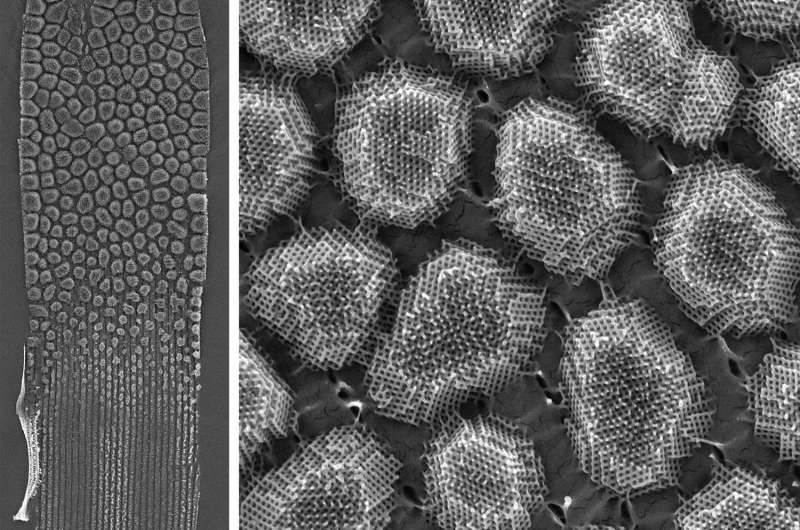
Images taken using scanning electron microscopy, high magnification light microscopy and x-ray nanotomography revealed gyroids with a definite size gradient—and which were not interconnected. From the perspective of moving along a scale from one end to the other, the gyroids on the surface grow larger, which suggested a dynamic growth process. This finding casts doubt on prior theories suggesting that the gyroids were generated from what has been described as a "pre-folded template."

The researchers suggest the structure they witnessed in one species is likely found in others, and claim their results add to the understanding of gyroid development, while also offering clues regarding how cellular structures develop in other creatures such as mitochondria or chloroplasts.

More information: Bodo D. Wilts et al. Butterfly gyroid nanostructures as a time-frozen glimpse of intracellular membrane development, Science Advances (2017). DOI: 10.1126/sciadv.1603119
Abstract
The formation of the biophotonic gyroid material in butterfly wing scales is an exceptional feat of evolutionary engineering of functional nanostructures. It is hypothesized that this nanostructure forms by chitin polymerization inside a convoluted membrane of corresponding shape in the endoplasmic reticulum. However, this dynamic formation process, including whether membrane folding and chitin expression are simultaneous or sequential processes, cannot yet be elucidated by in vivo imaging. We report an unusual hierarchical ultrastructure in the butterfly Thecla opisena that, as a solid material, allows high-resolution three-dimensional microscopy. Rather than the conventional polycrystalline space-filling arrangement, a gyroid occurs in isolated facetted crystallites with a pronounced size gradient. When interpreted as a sequence of time-frozen snapshots of the morphogenesis, this arrangement provides insight into the formation mechanisms of the nanoporous gyroid material as well as of the intracellular organelle membrane that acts as the template.
Journal information: Science Advances
© 2017 Phys.org


