Noninvasive magnetic resonance imaging of lung fibrogenesis with an amino acid targeted probe
Organs respond to injuries with the formation of new fibrous tissue, which can result in scarring. This process called fibrogenesis can now be monitored noninvasively on a molecular level, as American scientists report in the journal Angewandte Chemie. They have created a new gadolinium-based probe for magnetic resonance imaging that specifically reports the proteins involved in fibrogenesis. The imaging method may provide a quantitative assessment of the formation of the potentially harmful scar tissue.
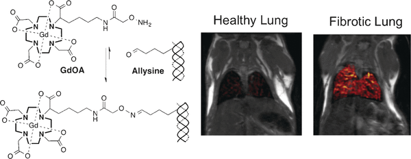
Natural wound healing and tissue injury involves the formation of collagen-based fibrous tissue to close the wound. In normal healing, the fibrous material is eventually replaced by normal tissue. If the injury is large or if the tissue is repeatedly damaged, the wound healing process may not be complete, resulting in scar formation that threatens the loss of function and even organ failure. On a molecular level, fibrogenesis is the accumulation and then remodeling of collagen, mainly by cross-linking, to create more rigid and tight structures. To monitor this process, Peter Caravan and collaborators from Massachusetts General Hospital and Harvard Medical School, USA, sought a molecular probe that could specifically recognize the components involved in fibrogenesis. They have created a functionalized gadolinium chelate as a probe for magnetic resonance imaging (MRI).
The probe was developed to target allysine, an amino acid indicative of active collagen cross-linking. "In active fibrogenesis, an active pool of allysine would be generated, but in stable disease or with therapeutic invention, these allysine moieties would be converted to cross-links," the authors reasoned. Thus, they designed an oxyamine-functionalized gadolinium chelate termed GdOA that would form stable oxime links with allysine and is "therefore expected to result in a strong MR signal enhancement on binding to allysine," as the authors proposed.
The GdOA meets the key criteria for MRI technology, as the researchers proved in numerous tests. It was stable, water-soluble, and excretable through the renal tract with very good pharmacokinetics, and it displayed high target selectivity in both test tube and in real mouse models. The scientists quantified pulmonary fibrogenesis in mice noninvasively by MRI using this probe, and they also monitored the suppression of fibrogenesis when the mice were treated with a specific medication. In this report, they studied fibrosis of the lung, but fibrogenesis is a common feature of many chronic diseases of the inner organs, and also in many cancers. This work gives a promising outlook to new specific MRI probes for disease detection, prognosis, and monitoring disease progression or treatment response across a range of human conditions.
More information: Philip A. Waghorn et al. Molecular Magnetic Resonance Imaging of Lung Fibrogenesis with an Oxyamine-Based Probe, Angewandte Chemie International Edition (2017). DOI: 10.1002/anie.201704773
Journal information: Angewandte Chemie , Angewandte Chemie International Edition
Provided by Wiley


