Miraculous proliferation

Bacteria able to shed their cell wall assume new, mostly spherical shapes. ETH researchers have shown that these cells, known as L-forms, are not only viable but that their reproductive mechanisms may even correspond to those of early life forms.
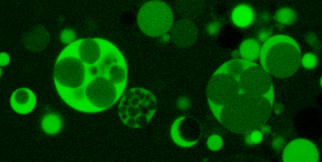
Researchers from a group led by ETH professor Martin Loessner discovered a few years ago that rod-shaped Listeria can become spherical. These L-forms differ fundamentally from normal forms of bacteria. They shed their cell wall, assume mostly spherical shapes and are capable of multiplying through budding-like processes in which the mother cell membranes form daughter vesicles. However, not all these vesicles actually contain genetic material.
L-forms may be created when cell wall-active antibiotics are used against Listeria, which can cause severe cases of food infection. The drugs cause the bacteria to shed their cell wall - the attack point for the medication. The resulting vesicles are enclosed only by a cytoplasmic membrane, which renders those antibiotics ineffective.
L-forms occur not only in Listeria, but have been described for many others bacteria. Moreover, Mycoplasma, Rickettsiae and Chlamydiae, all of which are human pathogens, also lack a stable cell wall. Yet, L-forms are viable only under certain conditions; if the osmotic conditions are not suitable or change rapidly, the cells become unstable and may burst.

Viable cell or dead product of nature?
Until now, researchers have not been able to clearly demonstrate viability of the resulting vesicles. But recent experiments by Loessner and his group have now show that L-forms are an independent form of life that can multiply indefinitely. "Our observations are not artefacts, but rather represent an alternative form of bacterial life," emphasises Loessner. The results of their latest study have just been published in the journal Nature Communications.
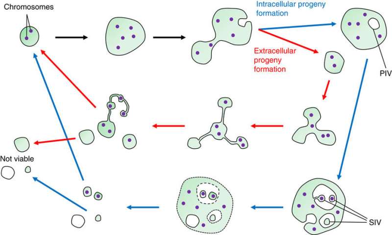
In their study, the researchers show that small membrane vesicles are formed by invagination into larger intracellular vesicles, thereby receiving cytoplasmic content and producing viable progeny.
When the cytoplasmic membrane of an L-Form cell invaginates into the cytoplasm, the mother cell produces a vesicle filled with substances from the surrounding medium. Consequently, this primary vesicle lacks cellular components and in particular the genetic material. However, by forming secondary vesicles through membrane extrusion into the primary ones these are filled with cytoplasmic material of the mother vesicle; which provides all the components of a cell needed for viability such as chromosomes and ribosomes, the production sites for proteins. Whether this second vesicle actually receives genetic material is, however, unregulated and random, but as a general rule provides enough volume and space that all vital processes can occur.
A crazy network of bubbles
Loessner considers it a very significant finding that in the course of their research on Listeria L-forms, they discovered small, elastic connector tubes between the outside vesicles, that look like "pearls on a chain". "The vesicles form a crazy network among themselves," he says. As intact membranes, the tubes consist of lipid molecules and they enable the vesicles to form a continuum, similar to fungal mycelium. Until they are fully separated, the vesicles can exchange cytoplasm via these tubes.
Similarly unusual is that in order to multiply, the L-forms require neither a cell wall nor the ring-forming FtsZ protein, which regular bacterial cells need to divide. "If we consider the early cells without rigid walls in the history of microbial evolution, then they most likely divided like the L-forms," explains Loessner. However, this is not a biological process but rather a physical one that depends directly on the amount of membrane material produced. "Multiplication follows the laws of thermodynamics." The L-forms may thus be compared to soap bubbles, which also owe their stability (and division) to purely physical principles.
More information: Patrick Studer et al, Proliferation of Listeria monocytogenes L-form cells by formation of internal and external vesicles, Nature Communications (2016). DOI: 10.1038/ncomms13631
Journal information: PLoS ONE , Nature Communications
Provided by ETH Zurich


