Researchers develop novel 3-D printing method for creating patient-specific medical devices

We've all seen pictures of premature babies in neonatal care units: tiny beings, some weighing just a bit over a pound, with plastic tubes snaking through their nose or mouth, or disappearing into veins or other parts of the body. Those tubes, or "catheters," are how the babies get the necessary oxygen, nutrients, fluid, and medications to stay alive. In the United States alone, nearly 500,000 premature babies are born each year.
The problem is, today's catheters only come in standard sizes and shapes, which means they cannot accommodate the needs of all premature babies. "With neonatal care, each baby is a different size, each baby has a different set of problems," says Randall Erb, assistant professor in the Department of Mechanical and Industrial Engineering. "If you can print a catheter whose geometry is specific to the individual patient, you can insert it up to a certain critical spot, you can avoid puncturing veins, and you can expedite delivery of the contents."
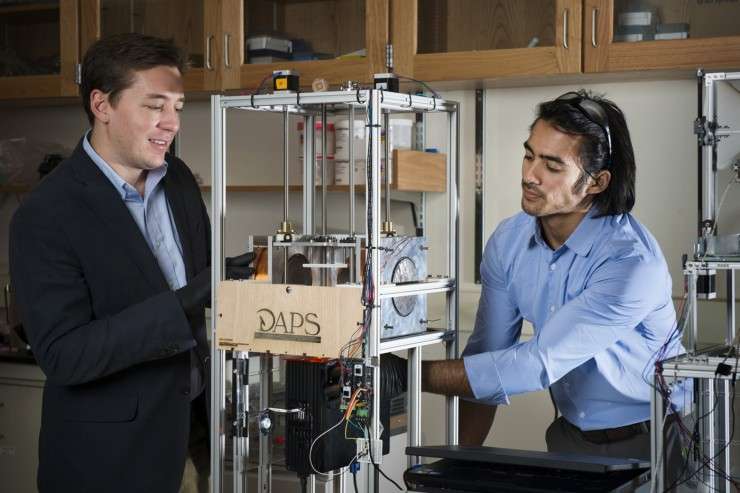
Erb's team has developed an innovative 3-D printing technology that uses magnetic fields to shape composite materials—mixes of plastics and ceramics—into patient-specific products. The biomedical devices they are developing, which include catheters, will be both stronger and lighter than current models and, with their customized design, ensure an appropriate fit. Their paper on the new technology appears in the Oct. 23 issue of Nature Communications.
Back to nature
Others have used composite materials in 3-D printing, says Joshua Martin, the doctoral candidate who helped design and run many of the experiments for the paper. What sets their technology apart, say Erb and Martin, is that it enables them to control how the ceramic fibers are arranged—and hence control the mechanical properties of the material itself.
That control is critical if you're crafting devices with complex architectures, such as customized miniature biomedical devices. Within a single patient-specific device, the corners, the curves, and the holes must all be reinforced by ceramic fibers arranged in just the right configuration to make the device durable. This is the strategy taken by many natural composites from bones to trees.
Consider the structure of human bone. Fibers of calcium phosphate, the mineral component of bone, are naturally oriented just so around the holes for blood vessels in order to ensure the bone's strength and stability, enabling, say, your femur to withstand a daily jog.
"We are following nature's lead," explains Martin, PhD'17, "by taking really simple building blocks but organizing them in a fashion that results in really impressive mechanical properties." Using magnets, Erb and Martin's 3-D printing method aligns each minuscule fiber in the direction that conforms precisely to the geometry of the item being printed.
"These are the sorts of architectures that we are now producing synthetically," says Erb, who has received a $225,000 Small Business Technology Transfer grant from the National Institutes of Health to develop the neonatal catheters with a local company. "Another of our goals is to use calcium phosphate fibers and biocompatible plastics to design surgical implants."
Magnetic attraction
The magnets are the defining ingredient in their 3-D printing technology. Erb initially described their role in the composite-making process in a 2012 paper in the journal Science.
First the researchers "magnetize" the ceramic fibers by dusting them very lightly with iron oxide, which, Martin notes, has already been FDA approved for drug-delivery applications. They then apply ultralow magnetic fields to individual sections of the composite material—the ceramic fibers immersed in liquid plastic—to align the fibers according to the exacting specifications dictated by the product they are printing.
In a video accompanying the Science article, you can see the fibers spring to attention when the magnetic field is turned on. "Magnetic fields are very easy to apply," says Erb. "They're safe, and they penetrate not only our bodies—think of CT scans—but many other materials."
Finally, in a process called "stereolithography," they build the product, layer by layer, using a computer-controlled laser beam that hardens the plastic. Each six-by-six inch layer takes a mere minute to complete.
"I believe our research is opening a new frontier in materials-science research," says Martin. "For a long time, researchers have been trying to design better materials, but there's always been a gap between theory and experiment. With this technology, we're finally scratching the surface where we can theoretically determine that a particular fiber architecture leads to improved mechanical properties and we can also produce those complicated architectures."
More information: Joshua J. Martin et al. Designing bioinspired composite reinforcement architectures via 3D magnetic printing, Nature Communications (2015). DOI: 10.1038/ncomms9641
R. M. Erb et al. Composites Reinforced in Three Dimensions by Using Low Magnetic Fields, Science (2012). DOI: 10.1126/science.1210822
Journal information: Nature Communications , Science
Provided by Northeastern University


