Unlocking secrets of how fossils form
Fossils tell amazing stories and inspire them, too—just think of this summer's "Jurassic World" blockbuster. But because some of the processes that preserve fossils are not well understood, there's still more information that they could reveal. Now scientists report in ACS' journal Analytical Chemistry a new way to probe fossils to find out how these ancient remains formed in greater detail than before.
When most organisms die, they biodegrade and leave little behind. But if they get trapped in sediments that harbor few bacteria and loads of dissolved minerals, they can become fossilized and preserved for millions of years. Scientists use a variety of techniques on the ancient specimens to determine details about lifestyles and diets, as well as information about the geographical distribution of the creatures. One of those methods called scanning electron microscopy, or SEM, showed particular promise for revealing new information about fossils. So Amauri J. Paula and colleagues expanded on this method.
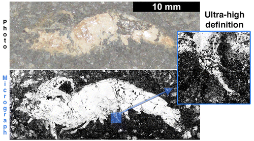
The researchers used a large-field SEM approach to analyze a shrimp fossil from the Araripe Basin, a place in northeastern Brazil known among paleontologists as a treasure trove of flying pterosaur remains. The shrimp specimen dates back to the Cretaceous period, when dinosaurs still roamed the planet. The technique provided evidence for the first time that a rare fossilization process occurred in the basin. It also showed that the fossil over millions of years developed a surprising fractal characteristic—a still-unexplained, repeating pattern most commonly recognized in snowflakes but also found in structures as large as spiral galaxies.
More information: Large-Field Electron Imaging and X-ray Elemental Mapping Unveil Morphology, Structure and Fractal Features of a Cretaceous Fossil at the Centimetre Scale, Anal. Chem., Article ASAP. DOI: 10.1021/acs.analchem.5b02815
Abstract
We used here a scanning electron microscopy approach that detected backscattered electrons (BSEs) and X-rays (from ionization processes) along a large-field (LF) scan, applied on a Cretaceous fossil of a shrimp (area ∼280 mm2) from the Araripe Sedimentary Basin. High-definition LF images from BSEs and X-rays were essentially generated by assembling thousands of magnified images that covered the whole area of the fossil, thus unveiling morphological and compositional aspects at length scales from micrometers to centimeters. Morphological features of the shrimp such as pleopods, pereopods, and antennae located at near-surface layers (undetected by photography techniques) were unveiled in detail by LF BSE images and in calcium and phosphorus elemental maps (mineralized as hydroxyapatite). LF elemental maps for zinc and sulfur indicated a rare fossilization event observed for the first time in fossils from the Araripe Sedimentary Basin: the mineralization of zinc sulfide interfacing to hydroxyapatite in the fossil. Finally, a dimensional analysis of the phosphorus map led to an important finding: the existence of a fractal characteristic (D = 1.63) for the hydroxyapatite–matrix interface, a result of physical-geological events occurring with spatial scale invariance on the specimen, over millions of years.
Journal information: Analytical Chemistry
Provided by American Chemical Society


