Numerically identifying pollen grains improves on conventional ID method

Researchers have developed a new quantitative – rather than qualitative – method of identifying pollen grains that is certainly nothing to sneeze at.
The research appears in the journal Proceedings of the Royal Society B: Biological Sciences.
Since the invention of the earliest light microscopes, the classification and identification of pollen and spores has been a highly subjective venture for those who use these tiny particles to study vegetation in their field, palynology.
However, according to the lead author of the study, Luke Mander, a former postdoctoral researcher in the laboratory of Illinois professor of plant biology Surangi Punyasena, the limitations imposed by these descriptive rather than numerical methods have kept researchers from classifying pollen and spores beyond a general level.
"Grass pollen classification is a long-standing problem in palynology," Mander said. "Many researchers have tried to classify these things by eye, looking down a standard light microscope and noting some differences in their sizes and other aspects of their morphology, but the differences between the species is so subtle."
Mander said he and his colleagues set out to find a less subjective and more numbers-based method of grass-pollen grain classification and identification.
"We decided to use a quantitative analysis because we wanted to move beyond the idea that we just use natural language to describe the morphology and differences between the species. We wanted to be able to put numbers on them to generate robust and repeatable classifications," Mander said.
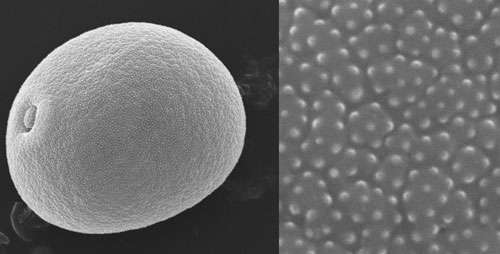
The researchers began by using scanning electron microscopes to capture detailed, high-resolution images of 12 different species of grass pollen. They measured the size and number of some of the pollen grains' morphological features, which, under the microscope, look like "little spines," according to Mander. The team, which included Florida State University mathematics professor Washington Mio and his graduate student Mao Li, then devised a novel method of quantifying the intricacies that exist among these species' spines.
"If you zoom in on an image it becomes very pixelated, and we thought of those pixels as little nodes in a network," Mander said. "We quantified the complexity of the networks that were formed by the pixels and used that measure of complexity to generate our classification."
This new method was 77.5 percent accurate, classifying 186 out of 240 specimens correctly. When seven human subjects were given the scanning electron microscopy images and a reference library, individually their classifications were 68.3 to 81.7 percent accurate. Between the subjects, however, there was relatively little classification consistency, as only 28.3 percent of the specimens were correctly classified by every subject.

"You can see the differences between species with your eyes reasonably well, but individual workers have very different classifications. In our analysis, we found that consistency between workers was very low, and that's the crucial difference. The quantitative analysis gives you much better repeatability and consistency," Mander said.
Punyasena said that her lab will continue to use these quantitative methods in their studies of the pollen fossil record.
"We are already working on a study of fossil spruce," she said. "We hope that texture can be used to tell apart four species, including one extinct spruce, and allow us to reconstruct the population dynamics of these species during the end of the last Ice Age in the southeastern U.S. This may explain why three of these species were able to migrate and establish northern populations, while the fourth species went extinct."
Punyasena also believes that those who conduct qualitative research in fields beyond palynology "from the description of fossil species to the recognition of mutant phenotypes in genetic research" will be able to apply the new approach to their work.
"Developing better, quantitative ways of describing biological shape and texture would allow many fields to establish more rigorous and consistent criteria for working with morphology and assessing morphological differences," Punyasena said.
More information: rspb.royalsocietypublishing.or … /1770/20131905.short
Journal information: Proceedings of the Royal Society B
Provided by University of Illinois at Urbana-Champaign


.jpg)


