A microscopic approach to the magnetic sensitivity of animals

Researchers at the University of Tokyo have succeeded in developing a new microscope capable of observing the magnetic sensitivity of photochemical reactions believed to be responsible for the ability of some animals to navigate in the Earth's magnetic field, on a scale small enough to follow these reactions taking place inside sub-cellular structures.
Several species of insects, fish, birds and mammals are believed to be able to detect magnetic fields - an ability known as magnetoreception. For example, birds are able to sense the Earth's magnetic field and use it to help navigate when migrating. Recent research suggests that a group of proteins called cryptochromes and particularly the molecule flavin adenine dinucleotide (FAD) that forms part of the cryptochrome, are implicated in magnetoreception. When cryptochromes absorb blue light, they can form what are known as radical pairs. The magnetic field around the cryptochromes determines the spins of these radical pairs, altering their reactivity. However, to date there has been no way to measure the effect of magnetic fields on radical pairs in living cells.
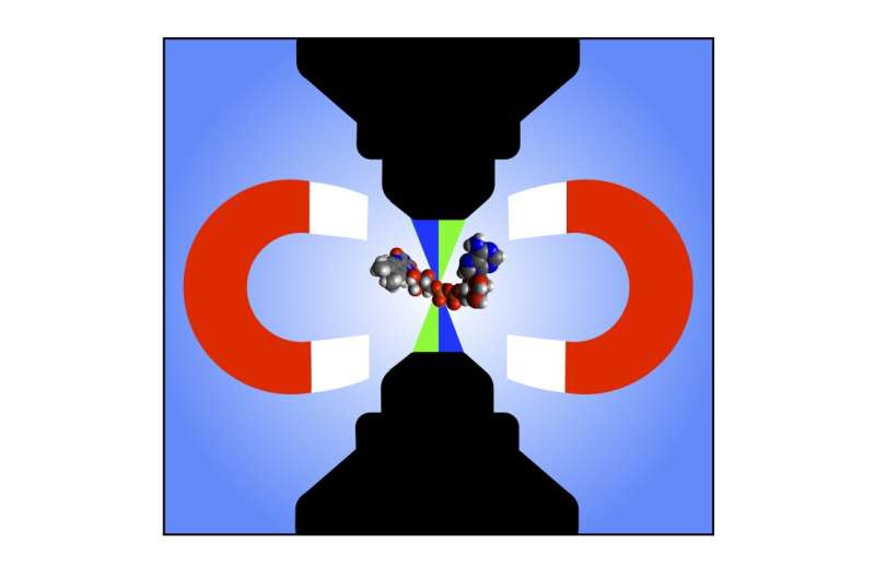
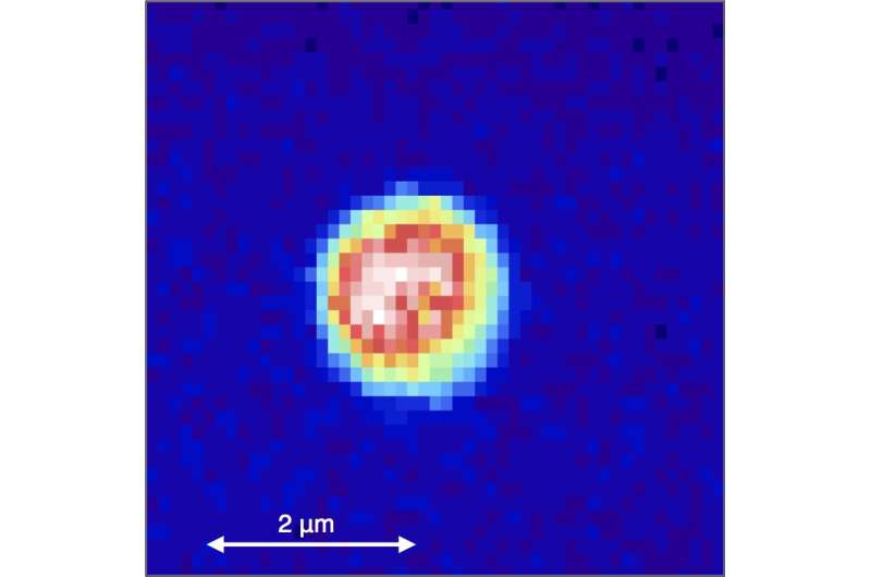
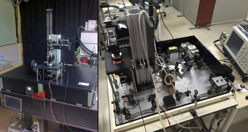
The research group of Associate Professor Jonathan Woodward at the Graduate School of Arts and Sciences are specialists in radical pair chemistry and investigating the magnetic sensitivity of biological systems. In this latest research, PhD student Lewis Antill made measurements using a special microscope to detect radical pairs formed from FAD, and the influence of very weak magnetic fields on their reactivity, in volumes less than 4 millionths of a billionth of a liter (4 femtoliters). This was possible using a technique the group developed called TOAD (transient optical absorption detection) imaging, employing a microscope built by postdoctoral research associate Dr. Joshua Beardmore based on a design by Beardmore and Woodward.

"In the future, using another mode of the new microscope called MIM (magnetic intensity modulation), also introduced in this work, it may be possible to directly image only the magnetically sensitive regions of living cells," says Woodward. "The new imaging microscope developed in this research will enable the study of the magnetic sensitivity of photochemical reactions in a variety of important biological and other contexts, and hopefully help to unlock the secrets of animals' miraculous magnetic sense."

More information: Joshua P. Beardmore, Lewis M. Antill, and Jonathan R. Woodward, "Optical Absorption and Magnetic Field Effect Based Imaging of Transient Radicals" Angewandte Chemie International Edition. Early View: 2015/6/3, DOI: 10.1002/anie.201502591
Journal information: Angewandte Chemie International Edition
Provided by University of Tokyo


