New imaging approach accurately measures lipid and metabolite distributions in biological samples

To understand how cells converse, scientists at Pacific Northwest National Laboratory and Oregon Health & Science University designed an approach that accurately determines the spatial location of molecules and quantifies lipids and metabolites in biological samples. The new approach efficiently accounts for signal suppression or matrix effect (see sidebar) that may significantly alter molecules' distributions obtained in mass spectrometry imaging experiments. Compensation for matrix effects is achieved by adding appropriate internal standards to the solvent used in nano-DESI imaging.
"This approach comes at no cost in terms of sample preparation and experiment design," said Dr. Julia Laskin, a physical chemist at PNNL who led the study and the development of nano-DESI imaging.
Scientists use mass spectrometry imaging to determine spatial distributions of molecules in diminutive samples, such as slices of tissue or microbial colonies. Unfortunately, the technique is prone to matrix effects, which can skew the results. The new technique compensates for the matrix effects and determines the real distribution of lipids and metabolites in the sample providing valuable information for cost-effective production of biofuels and pharmaceuticals.
"Our technique reflects what's in the sample," said Dr. Ingela Lanekoff, an analytical chemist who worked on the project. "This is a new way of gathering and analyzing data that will feed into a lot of biological applications in the future."
Dr. Susan Stevens and Mary Stenzel-Poore from Oregon Health & Science University acquired mouse brain tissue harvested after the mouse had undergone a stroke, technically known as a middle cerebral artery occlusion stroke model sample. This type of tissue was chosen because half the brain shows the chemical aftermath of the stroke and the other half remains almost intact.

"This gives you a control on one side of the tissue section and the sample affected by stroke on the other, which is very helpful for understanding matrix effects," said Laskin.
The team at PNNL placed thin sections of the tissue sample on a glass slide and analyzed them using nano-DESI imaging, which stands for Nanospray Desorption Electrospray Ionization Mass Spectrometry imaging. Developed in 2009, nano-DESI allows researchers to analyze small quantities of materials with minimal sample preparation. Researchers have used it to analyze particles found in the air, microbial communities, petroleum, and other complex samples.
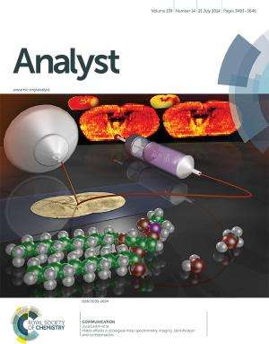
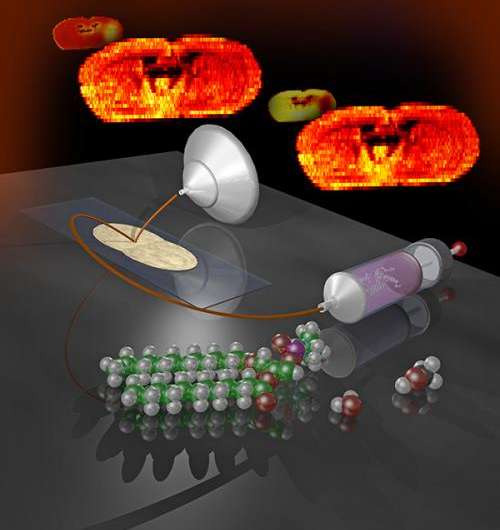
The key is a thin stream of liquid, called a liquid bridge. The bridge is built by pumping liquid through two glass capillaries positioned close to each other. When the liquid bridge touches the sample, it removes molecules from the sample surface. These molecules are then delivered in the solvent stream to a mass spectrometer inlet through the second capillary, which transforms it into charged droplets. Bare ions are generated from these droplets and detected by a mass spectrometer. Mass spectra obtained using this approach provide information on the type and abundance of hundreds of molecules extracted from the location where the drop was placed.
"We use a gentle extraction method," said Lanekoff.
To combat matrix effects, the scientists added a small amount of two lipid standards to the solvent usually used in nano-DESI. The solution containing known concentrations of standards was used in imaging experiments. By taking ratios of signal intensities of lipids extracted from the sample to lipid standards present in solution, they compensated for the matrix effects.
"We were able to distinguish what was really changing in the sample versus what was changing just because of the nature of the matrix effects," said Lanekoff.
Using this technique, the team identified and compensated for two types of matrix effects: one originating from differences in levels of sodium and potassium in the sample, and another one originating from differences in tissue composition. Molecular distributions free of matrix effects are markedly different from the raw ion images and reveal true variations in the distribution of phosphatidylcholines, a major component of biological membranes and an important class of signaling molecules supporting learning and memory processes in the brain.
The team is continuing to use nano-DESI in a broad range of applications in the biological community and beyond, including understanding metabolic exchange between microbial and fungal communities and identifying different cell types in healthy lung tissues with PNNL Laboratory Fellow Dr. Rick Corley.
More information: Lanekoff I, SL Stevens, MP Stenzel-Poore, and J Laskin. 2014. "Matrix Effects in Biological Mass Spectrometry Imaging: Identification and Compensation." Analyst 139:3528-3532. DOI: 10.1039/C4AN00504J
Journal information: Analyst
Provided by Pacific Northwest National Laboratory


