Researchers make proton radiation in cancerous tissue visible using ultrasound technology

Using ultrasound technology, physicists from the Munich-Centre for Advanced Photonics make proton radiation in cancerous tissue visible.
In future, the irradiation of tumors with protons could become more precise. Medical physicists from the Munich-Centre for Advanced Photonics (MAP) at the Ludwig-Maximilians-Universität (LMU), together with physicists from the Technical University (TUM), the Helmholtz Zentrum München (HMGU) and the Universität der Bundeswehr München (UniBWM) have combined conventional ultrasound technology with proton irradiation of a tumor. Using ionoacoustic technology they developed, they are able to observe the action of proton beams in real time via ultrasound.
A large number of tumors can be treated with radiation consisting of protons (positively charged hydrogen atoms), which attack and destroy the cancer cells of the tumor. However, it is crucial that the protons attack and kill only cancerous cells while sparing the surrounding healthy tissue. Doctors must therefore direct the energy of the protons precisely within the tumor in order to have maximum impact on the tumor cells.
In clinical applications, it is therefore important to know where the radiation from protons unleashes its maximum effect. In the human body, this is precisely where protons get stopped. This point of maximum dose delivery is known as the "Bragg peak" and should only occur within the tumor.
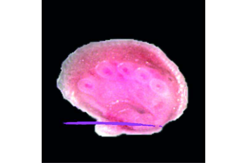
The medical physicists from the Munich-Centre for Advanced Photonics at LMU, in collaboration with research groups from TUM/HMGU and UniBWM, have now developed a method with which they can check where ion radiation deposits dose in a tumor during irradiation. The physicists have combined conventional ultrasound measurements with the simultaneous measurement of the ultrasonic signal caused by the proton irradiation. They first succeeded in making a beam of protons visible in tissue with an ultrasound image of this piece of tissue during a preclinical experiment. Using their own developed "ionoacoustic" technology, they are now able to track where ion radiation reaches its greatest effectiveness in the body – in real time, and in three dimensions. The researchers were thus able to determine the accuracy of the proton Bragg peak to within less than a millimeter. In addition, using controlled illumination with laser light, they were able to simultaneously measure an optoacoustic image of the irradiated tissue.
In order to adapt ionoacoustics for clinical practice, the physicists want to modify this ultrasound technology so that signals can be measured even at conventionally used levels of radiation at therapeutic energies.
At present, proton beams are still produced using large and expensive accelerator facilities. But new laser technologies being developed at the Munich-Centre for Advanced Photonics and its laser research center, the Centre for Advanced Laser Applications (CALA), promise cheaper – and possibly energetically better adapted – proton radiation for medical use. For laser-driven beam production, ionoacoustics promises to be a particularly suitable and highly accurate measurement method to make proton therapy more targeted and thus potentially more beneficial to patients in future.
More information: Stephan Kellnberger et al. Ionoacoustic tomography of the proton Bragg peak in combination with ultrasound and optoacoustic imaging, Scientific Reports (2016). DOI: 10.1038/srep29305
Journal information: Scientific Reports
Provided by Ludwig Maximilian University of Munich


