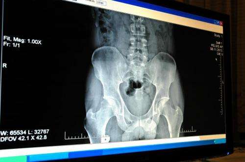
X-rays in five seconds
Wanting to replace the medical equipment for taking X-rays, the Mexican Society of Radiology (CMR) created a system of digital x-ray imaging, which replaces the traditional plaque by a solid detector, which delivers results in five seconds. Analog equipment take six minutes to develop the traditional film.
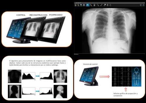
Engineer Nashelli Cuaranta Monroy, CMR researcher, explained that the system called ARiX RAD obtains X-ray images that aid in the diagnosis of various diseases, like identifying bone fractures, kidney stones or tumors.
The difference with this new equipment is that it replaces the radiographic film for a radiation detector, which will provide electrical signals proportional to the levels of radiation received. The digital detectors cointain a device called a cesium iodide scintillator, that converts X-rays into light, which in turn is converted into digital signals through a layer of amorphous silicon photodiodes that are processed to obtain a picture.
A digital image allows a more accurate assessment by using software tools that enable enhancement and post-processing of the radiograph that unfolds in conventional monitors. In addition, control and virtual management facilitates distribution through the internet, enabling processes like teleradiology and remote diagnostics.
The ARiX RAD system is integrated by a computer that performs three functions: first it selects the radiographic technique for the study; then it converts electrical signals from the radiation detector into a digital image, and finally comes the post-processing were processing and manipulating radiographs with special software occurs.
The system has an interface that allows to easily select the radiographic technique used and therefore the amount of milliamperes, kilovolts and the time required to perform an X-ray according to the anatomic area, projection and size of the patient.
To take an X-ray an intuitive interface (program design) is used, which graphically displays the different complexions of patients (infant, thin, normal, strong) and different projection (frontal view, lateral or oblique) and for each of the above shows the different anatomical regions which can be radiographed, explained the CMR engineer.
This system is easy to use because it allows an operator to obtain radiographic images with minimal training. Another benefit is that by using a processing algorithm to enhance the quality of image of the anatomical region radiographed, secondary radiation (noise) is filtered preventing disturbance in image quality.
With the implementation of the system manufacturing costs and maintenance are reduced, as well as the installation space and equipment which benefit patients by making them wait less time.
Provided by Investigación y Desarrollo


