Scientist uses state-of-the-art microscopy to discover drug candidates for cancer
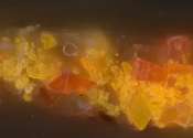
Microscopy has been making leaps and bounds in recent years. Science that was inconceivable a few years ago has become a matter of programming state-of-the-art microscopes to process reams of data. Dr. Gabriel Frank quickly ...