This article has been reviewed according to Science X's editorial process and policies. Editors have highlighted the following attributes while ensuring the content's credibility:
fact-checked
peer-reviewed publication
proofread
New 3D anatomical atlas of the African clawed frog increases understanding of development and metamorphosis processes

A 3D anatomical atlas of the model organism Xenopus laevis (the African clawed frog) is now available to aid researchers in understanding embryonic development and metamorphosis—the intriguing process by which a tadpole transforms into a mature frog.
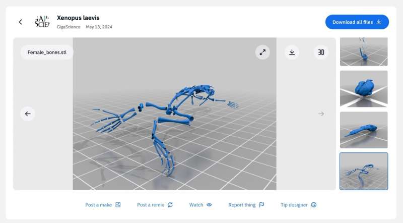
The lack of availability of this type of data has greatly limited the ability to assess and understand these complex processes. To increase access and interactivity for researchers, science educators and even 3D printing enthusiasts, this data has been converted into freely available embeddable digital files for 3D-viewing with Sketchfab and as 3D printing files available on Thingiverse. This work, along with all the available data, has been published in the journal GigaScience.
The African clawed frog (Xenopus laevis) has become a well-understood and versatile vertebrate model organism for studies in developmental biology and other disciplines due to the availability of multiple types of data, from foundational transplantation experiments for the field of embryology in the early twentieth century to present day experiments using high-quality genome sequencing technology.
This easy-to-breed frog is particularly suited to studies of body plan reorganization during the big changes that happen when the tadpole transforms into a mature frog, a process called metamorphosis. However, to move forward in gaining greater understanding of these processes, there is a great need for an additional type of data.
Dr. Jakub Harnos from Masaryk University (Czech Republic), a lead scientist of the study, explains that "a notable gap exists in the availability of comprehensive datasets encompassing Xenopus's late developmental stages."
To fill this void, the team of researchers now provide this missing data. The authors used X-ray microtomography, a 3D imaging technique, to create an anatomical atlas to more accurately describe the multiple stages of X. laevis development. Using detailed analysis of their 3D reconstructions at the various stages of development, the authors could pinpoint key changes that occur during the anatomical transformations at the stages from tadpole to froglet to mature adult.
One striking example of the shape changes that can be tracked in great detail with this new high-resolution data is the adjustment of the position of the developing frog's eyes, and the exact timing of this shift. With advancing development, the distance between the eyes progressively decreases.
"This adaptation aligns well with the frog's life strategy, transitioning from a water-dwelling tadpole with lateral eyes to an adult with eyes positioned on top of the head for a submerged lifestyle, reminiscent of crocodilians," the authors note.
The frog's gut also undergoes significant remodeling during metamorphosis. Over an 8-day period, the intestine shortens by around 75%, and the coiling pattern changes drastically. This process, which is difficult to study with other methods, can be followed in fine detail by X-ray microtomography the researchers have produced.
Other anatomical facts that are showcased in high spatial and temporal resolution by the new 3D atlas are seeing the differences between male and female frogs (females end up larger, overall) and the very subtle positioning of the teeth of X. laevis, which are hidden behind the maxillary arch.
"Our study provides all X-ray microtomography data openly, allowing other researchers to investigate both soft and hard tissues in unprecedented detail in this key vertebrate model," Dr. Harnos emphasizes.
To enable scientists, educators and the 3D printing community easy access to printable models, a collection of 40 surface-rendered 3D models from the Xenopus laevis anatomical atlas are available on the design platform Thingiverse. Embeddable digital models can also be downloaded from the Sketchfab website and are viewable in the research paper.
More information: Jakub Harnos et al, Unveiling Vertebrate Development Dynamics in Frog Xenopus laevis using Micro-CT Imaging, GigaScience (2024). DOI: 10.1093/gigascience/giae037
Journal information: GigaScience
Provided by GigaScience


