This article has been reviewed according to Science X's editorial process and policies. Editors have highlighted the following attributes while ensuring the content's credibility:
fact-checked
peer-reviewed publication
trusted source
proofread
From growing roots, clues to how stem cells decide their fate

It might look like a comet or a shooting star, but this time-lapse video is actually a tiny plant root, not much thicker than a human hair, magnified hundreds of times as it grows under the microscope.
Researchers at Duke University have been making such movies by peering at stem cells near the root's tip and taking snapshots as they divide and multiply over time, using a technique called light sheet microscopy.
The work offers more than a front-row seat to the drama of growing roots. By watching how the cells divide in response to certain chemical signals, the team is finding new clues to how stem cells choose one developmental path over another.
The research could also point to new ways to prevent stem cell division from going awry, as happens in cancer and other diseases.
The team's latest results appeared Jan. 31 in the journal Nature.
The work touches on a fundamental question in biology, said associate research professor Cara Winter: "How do cells acquire their identities?" In other words, "how do you get all of the various cell types that make up an organism?"
Just as the human body is made up of many different kinds of cells—in the brain, muscles, bones and elsewhere—plants, too, contain various cell types specialized for different tasks.
Whether in the roots, branches, flowers or leaves, virtually all tissues of a plant descend from small groups of unspecialized stem cells that produce new cells by dividing.
Each time a stem cell divides, it faces a choice: it can either produce two new stem cells like itself, or it can make one copy of itself plus one cell that will branch off to become something new.
It's the latter process, known as asymmetric division, that generates the myriad cell types needed to form a complex organism like a plant, or a human being.
An obvious question then is: How do dividing stem cells choose one path over the other?
This was the question driving Winter and co-first author Pablo Szekely, both researchers in the lab of late biologist Philip Benfey of Duke, as they watched days of root growth in Arabidopsis thaliana, a spindly member of the mustard family.
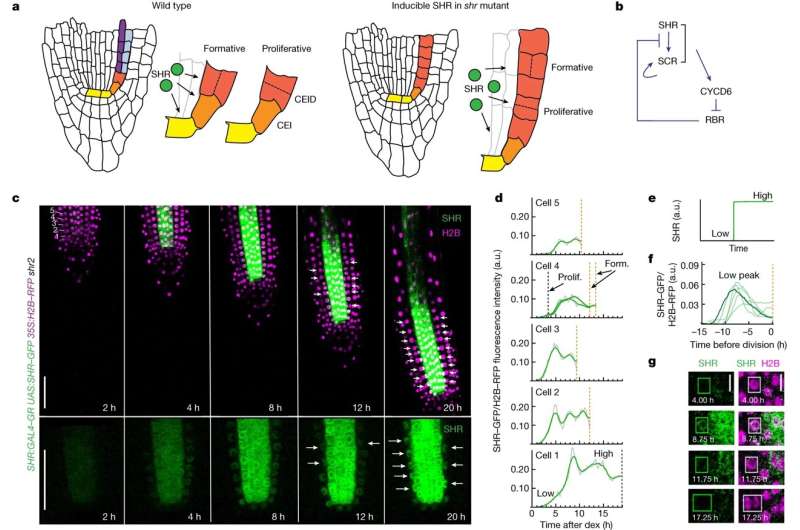
The researchers focused on two key regulators of cell division in Arabidopsis—proteins called short-root and scarecrow that, together, prompt dividing root cells to make the switch.
By labeling these proteins with glowing fluorescent tags, they were able to track the activity of the proteins and their effects on dividing stem cells in real time. Light-sheet microscopy allowed them to peer inside the roots' translucent tissues for up to 50 hours without harming them.
Counter to previous predictions, the researchers showed that even low levels of these proteins, present early in the process of one cell becoming two, are enough to trigger a switch to asymmetric division.
"All they have to do is reach a certain threshold," said Szekely, who joined the Benfey lab as a postdoctoral researcher in 2020.
The findings have implications for humans and other animals too, the researchers said.
That's because, although plants and animals diverged more than a billion years ago, they inherited much of the same basic molecular tool kit—including many of the same "housekeeping" genes that are necessary for cells to function.
The same genes that regulate cell division in plants like Arabidopsis perform similar jobs in animals, including humans. Previous research shows that when asymmetric division is disrupted, cells can multiply out of control and form tumors.
"Cells need to have a program during development: first divide like this, then divide like that," Szekely said. "It has to be tightly regulated in order for everything to work."
More information: Cara M. Winter et al, SHR and SCR coordinate root patterning and growth early in the cell cycle, Nature (2024). DOI: 10.1038/s41586-023-06971-z
Journal information: Nature
Provided by Duke University


